What Does Each Part Of A Microscope Do ?
Each part of a microscope has a specific function. The eyepiece, or ocular lens, is where the viewer looks through to observe the specimen. The objective lenses, located on a rotating nosepiece, provide different levels of magnification. The stage is where the specimen is placed for observation. The condenser focuses and directs light onto the specimen. The diaphragm controls the amount of light passing through the condenser. The light source, typically a lamp, provides illumination for the specimen. The coarse and fine focus knobs are used to adjust the focus of the microscope. Finally, the arm and base provide support and stability to the microscope.
1、 Eyepiece: Provides magnification for viewing the specimen.
Each part of a microscope plays a crucial role in the overall functioning of the instrument. The eyepiece, also known as the ocular lens, is one of the key components of a microscope. Its primary function is to provide magnification for viewing the specimen.
The eyepiece is located at the top of the microscope and is the part through which the viewer looks to observe the specimen. It typically contains a lens or a set of lenses that magnify the image produced by the objective lens. The magnification power of the eyepiece can vary depending on the microscope model, but it is commonly 10x.
When light passes through the objective lens and the specimen, it forms an image that is then magnified by the eyepiece. This allows the viewer to see the specimen in greater detail and clarity. The eyepiece also helps to focus the image, ensuring that it appears sharp and clear to the viewer.
In recent years, advancements in technology have led to the development of digital microscopes, which incorporate a camera in the eyepiece. This allows for the capture of images and videos of the specimen, which can be viewed on a computer or other digital devices. This digital integration has revolutionized microscopy, enabling easier sharing of observations and facilitating analysis and documentation.
In conclusion, the eyepiece of a microscope provides the necessary magnification for viewing the specimen. It is an essential component that allows scientists, researchers, and students to observe and study microscopic structures with greater clarity and precision.

2、 Objective Lens: Gathers light and magnifies the specimen.
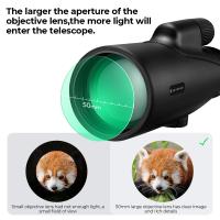
Each part of a microscope plays a crucial role in the overall functioning of the instrument. One of the key components is the objective lens, which is responsible for gathering light and magnifying the specimen. The objective lens is located at the lower end of the microscope and is usually available in different magnification powers, such as 4x, 10x, 40x, and 100x.
The primary function of the objective lens is to collect light from the specimen and focus it onto the intermediate image plane. This allows for a clear and magnified view of the specimen. The magnification power of the objective lens determines the level of detail that can be observed. For example, a 10x objective lens will magnify the specimen 10 times larger than its actual size.
In recent years, advancements in microscope technology have led to the development of high-quality objective lenses with improved resolution and clarity. These lenses are designed to minimize aberrations and enhance image quality. Additionally, some objective lenses are equipped with special coatings to reduce glare and improve contrast.
Objective lenses are typically mounted on a rotating nosepiece, allowing the user to easily switch between different magnification powers. This versatility is essential for examining specimens at various levels of detail.
In conclusion, the objective lens of a microscope is a critical component that gathers light and magnifies the specimen. It enables scientists, researchers, and students to observe microscopic structures with enhanced clarity and detail. Ongoing advancements in objective lens technology continue to push the boundaries of microscopic imaging, allowing for even more precise and accurate observations.

3、 Stage: Supports the specimen for observation.
Each part of a microscope plays a crucial role in the observation and analysis of specimens. One of the key components is the stage, which supports the specimen for observation. The stage is a flat platform that holds the specimen in place and allows for precise positioning and movement. It typically includes clips or mechanical holders to secure the specimen in place.
The stage is designed to provide stability and control during the observation process. It allows the user to manipulate the specimen and adjust its position to achieve the desired focus and clarity. By providing a stable platform, the stage ensures that the specimen remains in the field of view and does not move or shift during observation.
In addition to supporting the specimen, the stage often includes mechanical controls for precise movement. These controls allow the user to move the specimen horizontally (x-axis) and vertically (y-axis) to navigate across the slide and explore different areas of interest. Some advanced microscopes even offer motorized stages that can be controlled electronically, providing even greater precision and ease of use.
The stage is an essential part of the microscope as it enables the user to position the specimen accurately and manipulate it for detailed examination. Without a stable and adjustable stage, it would be challenging to focus on specific areas of interest or conduct precise measurements. Therefore, the stage is a critical component that contributes to the overall functionality and effectiveness of a microscope.
In recent years, there have been advancements in stage technology, such as the development of automated stages that can be programmed to move the specimen automatically. These automated stages are particularly useful in fields like pathology and research, where large numbers of specimens need to be analyzed quickly and efficiently. They allow for high-throughput imaging and data collection, saving time and improving productivity.
Overall, the stage of a microscope is responsible for supporting the specimen and providing stability and control during observation. It is an essential component that enables precise positioning and movement, allowing for detailed examination and analysis of specimens.

4、 Condenser: Focuses light onto the specimen.
Each part of a microscope plays a crucial role in the overall functioning of the instrument. The condenser, in particular, is responsible for focusing light onto the specimen, ensuring optimal illumination and clarity during observation.
The condenser is located beneath the stage and consists of a series of lenses that work together to concentrate and direct light onto the specimen. By adjusting the position of the condenser, the amount and angle of light can be controlled, allowing for enhanced visualization of the specimen.
The primary function of the condenser is to increase the numerical aperture (NA) of the microscope system. The NA determines the resolving power of the microscope, which is the ability to distinguish fine details and structures. By focusing light onto the specimen, the condenser increases the NA, resulting in improved resolution and image quality.
In addition to focusing light, the condenser also plays a role in controlling contrast. By adjusting the aperture diaphragm, which is located within the condenser, the amount of light passing through the specimen can be regulated. This helps to enhance contrast and highlight specific features of interest.
It is important to note that the role of the condenser may vary depending on the type of microscope being used. For example, in a brightfield microscope, the condenser is essential for providing even illumination across the specimen. In contrast, in a phase contrast or darkfield microscope, the condenser is used to manipulate the light in a way that enhances contrast and visibility of specific structures.
In recent years, advancements in microscope technology have led to the development of more sophisticated condensers. These include features such as adjustable aperture diaphragms, iris diaphragms, and even automated condensers that can optimize illumination settings based on the specimen being observed. These advancements have further improved the functionality and versatility of the condenser, allowing for more precise and detailed observations.
In conclusion, the condenser is a critical component of a microscope that focuses light onto the specimen, enhancing resolution, contrast, and overall image quality. Its role in controlling illumination and optimizing visualization has made it an indispensable part of modern microscopy.


