What Is A Light Microscope Definition ?
A light microscope, also known as an optical microscope, is a scientific instrument that uses visible light and a system of lenses to magnify and observe small objects or specimens. It is one of the most commonly used types of microscopes in various fields of science, including biology, medicine, and materials science. The basic principle of a light microscope involves passing light through the specimen, which interacts with the sample and produces an image that can be viewed through the eyepiece or captured using a camera. The lenses in the microscope system help to magnify the image, allowing for detailed examination of the specimen's structure and morphology. Light microscopes can have different configurations, such as compound microscopes with multiple lenses or stereo microscopes for three-dimensional observation. They have played a crucial role in advancing scientific knowledge and understanding of the microscopic world.
1、 Optical principles and components of a light microscope.
A light microscope, also known as an optical microscope, is a scientific instrument that uses visible light and a series of lenses to magnify and observe small objects or organisms that are otherwise invisible to the naked eye. It is one of the most commonly used tools in biology and other scientific fields.
The optical principles of a light microscope involve the interaction of light with the specimen being observed. When light passes through the specimen, it undergoes refraction, which causes the light rays to bend. This bending of light allows the microscope to magnify the image of the specimen. The lenses in the microscope, including the objective lens and the eyepiece, work together to focus and magnify the image.
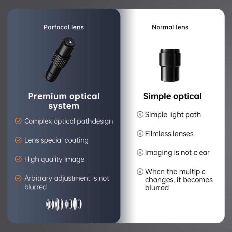
The components of a light microscope typically include an illuminator, which provides a light source, a condenser, which focuses the light onto the specimen, an objective lens, which magnifies the image, and an eyepiece, which further magnifies the image for the observer. The microscope also has a stage where the specimen is placed and a focus adjustment mechanism to bring the specimen into sharp focus.
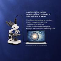
In recent years, advancements in technology have led to the development of more sophisticated light microscopes, such as confocal microscopes and fluorescence microscopes. These instruments utilize advanced optical techniques and components to enhance the resolution and contrast of the images obtained. Additionally, digital imaging and computer software have allowed for the capture, analysis, and storage of microscope images, enabling further research and analysis.
Overall, the light microscope remains an essential tool in scientific research, allowing scientists to explore the intricate details of the microscopic world and gain a deeper understanding of biological processes and structures.

2、 Types of light microscopes: compound, stereo, and phase contrast.
A light microscope is a type of microscope that uses visible light to magnify and observe small objects or organisms. It consists of a series of lenses that focus the light onto the specimen, allowing for detailed examination. The light microscope is one of the most commonly used tools in biological research and is essential for studying cells, tissues, and microorganisms.
There are several types of light microscopes, including compound, stereo, and phase contrast microscopes. Compound microscopes are the most widely used and have two sets of lenses - the objective lens and the eyepiece. They provide high magnification and resolution, allowing for the observation of fine details in cells and tissues.
Stereo microscopes, also known as dissecting microscopes, are used for viewing larger specimens in three dimensions. They have lower magnification but provide a wider field of view, making them suitable for examining objects such as insects, plants, or geological samples.
Phase contrast microscopes are designed to enhance the contrast of transparent specimens, such as living cells, by converting differences in refractive index into variations in brightness. This technique allows for the visualization of cellular structures that would otherwise be difficult to observe.
In recent years, advancements in technology have led to the development of more sophisticated light microscopes, such as confocal microscopes and super-resolution microscopes. These instruments offer even higher resolution and imaging capabilities, allowing scientists to study biological processes at the molecular level.
Overall, light microscopes have revolutionized the field of biology by enabling scientists to explore the intricate world of cells and microorganisms. They continue to be an indispensable tool for research and discovery in various scientific disciplines.

3、 Magnification and resolution in light microscopy.
A light microscope is a type of microscope that uses visible light to magnify and resolve the details of a specimen. It consists of a series of lenses that work together to enlarge the image of the specimen, allowing scientists to observe and study its structure and characteristics.
Magnification refers to the ability of the microscope to make the specimen appear larger than its actual size. This is achieved by using a combination of objective lenses with different magnification powers. The total magnification is calculated by multiplying the magnification of the objective lens by the magnification of the eyepiece.
Resolution, on the other hand, refers to the ability of the microscope to distinguish between two closely spaced objects as separate entities. It is determined by the wavelength of the light used and the numerical aperture of the objective lens. The numerical aperture is a measure of the lens' ability to gather light and is influenced by the refractive index of the medium between the lens and the specimen.
In light microscopy, the maximum resolution is limited by the diffraction of light waves. According to the Abbe diffraction limit, the resolution is approximately half the wavelength of the light used. However, recent advancements in microscopy techniques, such as super-resolution microscopy, have pushed the limits of resolution beyond the diffraction limit. These techniques utilize fluorescent dyes and complex imaging algorithms to achieve resolutions at the nanometer scale, allowing scientists to observe cellular structures and processes in unprecedented detail.
In summary, magnification and resolution are key parameters in light microscopy. While magnification allows for larger images of the specimen, resolution determines the level of detail that can be observed. Ongoing advancements in microscopy techniques continue to enhance the capabilities of light microscopy, enabling scientists to explore the microscopic world with greater precision and clarity.

4、 Sample preparation techniques for light microscopy.
A light microscope is a type of microscope that uses visible light to magnify and observe small objects or samples. It is a widely used tool in various scientific fields, including biology, medicine, and materials science. The light microscope definition refers to the use of lenses and light sources to produce magnified images of samples.
Sample preparation techniques for light microscopy involve a series of steps to ensure that the sample is properly prepared for observation under the microscope. These techniques aim to enhance the visibility and contrast of the sample, allowing for better resolution and analysis.
The first step in sample preparation is fixation, which involves preserving the sample's structure and preventing decay or degradation. This is typically done by using chemical fixatives such as formaldehyde or glutaraldehyde. After fixation, the sample may undergo dehydration, embedding, and sectioning processes, depending on the nature of the sample and the desired analysis.
Staining is another crucial step in sample preparation for light microscopy. Stains are used to selectively color different components of the sample, making them more visible under the microscope. Various types of stains are available, including dyes that bind to specific cellular structures or fluorescent probes that emit light when excited by specific wavelengths.
In recent years, there have been advancements in sample preparation techniques for light microscopy. For example, immunostaining techniques have become more sophisticated, allowing for the visualization of specific proteins or molecules within a sample. Additionally, the development of super-resolution microscopy techniques has enabled researchers to achieve higher resolution and better visualization of subcellular structures.
Overall, sample preparation techniques for light microscopy play a crucial role in obtaining clear and detailed images of samples. These techniques continue to evolve, driven by advancements in technology and the need for more precise and accurate analysis in various scientific disciplines.


There are no comments for this blog.