What Is A Microscope Diaphragm ?
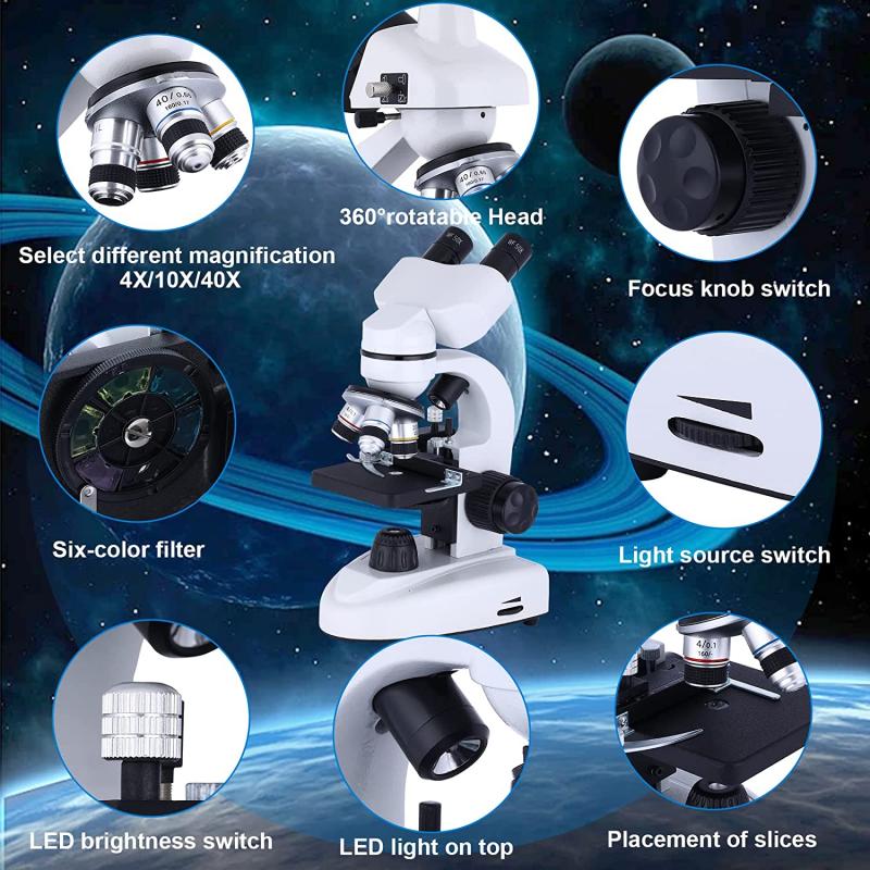
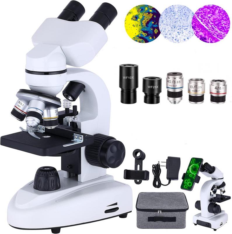
A microscope diaphragm is a device located beneath the stage of a microscope that controls the amount of light passing through the specimen. It consists of a circular or rectangular aperture with adjustable openings of different sizes. By adjusting the diaphragm, the user can regulate the intensity and angle of the light illuminating the specimen. This helps in achieving optimal contrast and resolution during microscopic observations. The diaphragm is an essential component in microscopy as it allows the user to control the amount of light reaching the specimen, thereby enhancing the visibility of the details under examination.
1、 Definition and Function of a Microscope Diaphragm
A microscope diaphragm is a device located beneath the stage of a microscope that controls the amount of light passing through the specimen. It consists of a rotating disk with different-sized apertures or openings, allowing the user to adjust the intensity and focus of the light source. By altering the size of the diaphragm opening, the amount of light reaching the specimen can be regulated, resulting in improved image quality and clarity.
The main function of a microscope diaphragm is to control the illumination of the specimen. By adjusting the diaphragm, the user can optimize the contrast and brightness of the image, making it easier to observe and analyze the specimen. This is particularly important when dealing with specimens that are transparent or have low contrast.
In addition to controlling illumination, the diaphragm also plays a role in depth of field. By adjusting the size of the diaphragm opening, the user can manipulate the depth of field, which refers to the range of focus in an image. This can be useful when examining specimens with varying depths, allowing the user to focus on specific areas of interest.
It is worth noting that with advancements in microscope technology, some modern microscopes now come equipped with automated diaphragms. These automated diaphragms can adjust the aperture size based on the specific requirements of the specimen or the imaging technique being used. This allows for more precise control over the illumination and depth of field, enhancing the overall imaging capabilities of the microscope.
In conclusion, a microscope diaphragm is a crucial component that controls the amount of light passing through the specimen, optimizing image quality and clarity. It allows for the adjustment of illumination and depth of field, enabling users to observe and analyze specimens more effectively.
2、 Types of Microscope Diaphragms and Their Features
A microscope diaphragm is an adjustable device located beneath the stage of a microscope that controls the amount of light passing through the specimen. It consists of a series of adjustable apertures or openings that can be adjusted to regulate the intensity and direction of the light source.
There are several types of microscope diaphragms, each with its own unique features. The most common types include the iris diaphragm, the disk diaphragm, and the pinhole diaphragm.
The iris diaphragm is the most versatile and widely used type. It consists of a series of overlapping metal blades that can be adjusted to control the size of the aperture. This allows for precise control of the amount of light passing through the specimen, resulting in improved image quality and contrast.
The disk diaphragm, on the other hand, consists of a series of different-sized holes or apertures that can be rotated into position. This type of diaphragm is less versatile than the iris diaphragm but is still effective in controlling the amount of light.
The pinhole diaphragm is a specialized type of diaphragm that consists of a small hole or aperture. It is used primarily in confocal microscopy, where it helps to eliminate out-of-focus light and improve image resolution.
In recent years, there has been a growing interest in the development of advanced diaphragm technologies. For example, some researchers are exploring the use of liquid crystal diaphragms, which can be electronically controlled to provide even greater flexibility and precision in light control.
Overall, the microscope diaphragm plays a crucial role in controlling the amount and quality of light that reaches the specimen, ultimately influencing the clarity and resolution of the observed image.
3、 Adjusting the Microscope Diaphragm for Proper Illumination
A microscope diaphragm is a device located beneath the stage of a microscope that controls the amount of light passing through the specimen. It consists of a rotating disk with different-sized apertures or openings that can be adjusted to regulate the intensity and quality of the illumination.
The main purpose of the microscope diaphragm is to optimize the lighting conditions for observing the specimen. By adjusting the diaphragm, the user can control the amount of light that reaches the specimen, which is crucial for achieving proper illumination. This is particularly important when dealing with specimens that are transparent or have low contrast.
To adjust the microscope diaphragm for proper illumination, one needs to consider the type of specimen being observed and the objective lens being used. Generally, a smaller aperture is used for higher magnification objectives, as it helps to increase contrast and reduce glare. On the other hand, a larger aperture is suitable for lower magnification objectives, as it allows more light to pass through and provides a brighter image.
It is worth noting that the latest point of view regarding microscope diaphragms emphasizes the importance of using the appropriate aperture size to achieve optimal image quality. Additionally, advancements in microscope technology have led to the development of automated diaphragms that can adjust themselves based on the objective lens in use, further simplifying the process for users.
In conclusion, a microscope diaphragm is a crucial component that allows users to control the amount of light passing through the specimen. Adjusting the diaphragm for proper illumination is essential for obtaining clear and detailed images, and the latest advancements in this area have made the process even more user-friendly.

4、 Importance of the Microscope Diaphragm in Image Contrast
A microscope diaphragm is a device located beneath the stage of a microscope that controls the amount and angle of light that passes through the specimen. It consists of a series of adjustable apertures or openings that can be adjusted to regulate the intensity and direction of light. By manipulating the diaphragm, the user can optimize the image contrast and clarity.
The importance of the microscope diaphragm in image contrast cannot be overstated. It plays a crucial role in controlling the amount of light that reaches the specimen. By adjusting the diaphragm, the user can increase or decrease the intensity of light, which directly affects the contrast of the image. This is particularly important when observing specimens with low contrast, such as transparent or unstained samples.
Furthermore, the diaphragm also helps in controlling the angle of light that passes through the specimen. By adjusting the aperture size, the user can control the numerical aperture (NA) of the microscope, which determines the resolution and depth of field. A smaller aperture increases the depth of field, allowing more of the specimen to be in focus, while a larger aperture increases the resolution, enabling finer details to be observed.
In recent years, there has been a growing emphasis on the importance of optimizing image contrast in microscopy. Researchers have been exploring new techniques and technologies to enhance contrast, such as phase contrast, differential interference contrast, and darkfield microscopy. However, the microscope diaphragm remains a fundamental tool in achieving optimal image contrast. Its precise control over the amount and angle of light is essential for obtaining clear and detailed images, especially in challenging specimens.
In conclusion, the microscope diaphragm is a critical component for controlling image contrast in microscopy. Its ability to regulate the intensity and direction of light allows users to optimize the clarity and visibility of specimens. Despite advancements in contrast-enhancing techniques, the diaphragm remains an indispensable tool for achieving high-quality microscopy images.
