What Is A Transmission Electron Microscope ?
A transmission electron microscope (TEM) is a type of microscope that uses a beam of electrons to create highly detailed images of the internal structure of a specimen. It works by passing a beam of electrons through a thin section of the specimen, which interacts with the electrons to produce an image. The image is then magnified and focused onto a fluorescent screen or a digital detector, allowing the user to observe the specimen at a very high resolution. TEMs are capable of achieving magnifications up to several million times, revealing the fine details of the specimen's internal structure. They are widely used in various scientific fields, including materials science, biology, and nanotechnology, to study the morphology, composition, and behavior of samples at the atomic and molecular level.
1、 Principle of Transmission Electron Microscopy
A transmission electron microscope (TEM) is a powerful imaging tool used in scientific research to visualize the ultrastructure of materials at the atomic level. It operates on the principle of transmitting a beam of electrons through a thin specimen, which interacts with the specimen and produces an image on a fluorescent screen or a digital detector.
The TEM consists of several key components, including an electron source, electromagnetic lenses, a specimen holder, and a detector. The electron source emits a beam of electrons, which is then focused and accelerated by the electromagnetic lenses. The beam passes through the thin specimen, and the electrons that interact with the specimen are scattered or absorbed. The remaining electrons form an image that is magnified and projected onto a screen or captured by a detector.
The TEM offers several advantages over other microscopy techniques. It provides extremely high resolution, allowing scientists to observe structures as small as a few angstroms. It also offers a large depth of field, enabling the visualization of both surface and internal structures of the specimen. Additionally, the TEM can provide information about the chemical composition of the specimen through techniques such as energy-dispersive X-ray spectroscopy.
In recent years, advancements in TEM technology have further enhanced its capabilities. For example, aberration correction techniques have significantly improved the resolution and image quality. Additionally, the development of environmental TEMs allows for the observation of materials under controlled environmental conditions, such as high temperatures or gas atmospheres.
Overall, the transmission electron microscope is a vital tool in various scientific disciplines, including materials science, nanotechnology, biology, and chemistry. Its ability to provide detailed information about the structure and composition of materials at the atomic level continues to drive advancements in scientific research and technological innovation.
2、 Components and Instrumentation of Transmission Electron Microscope
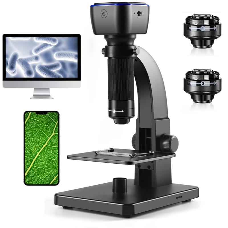
A transmission electron microscope (TEM) is a powerful scientific instrument used to study the structure and composition of materials at the atomic and molecular level. It utilizes a beam of electrons instead of light to image the specimen, allowing for much higher resolution and magnification compared to traditional light microscopes.
The basic components of a TEM include an electron source, electromagnetic lenses, a specimen holder, and a detector. The electron source, typically a heated tungsten filament or a field emission gun, emits a beam of electrons. The electromagnetic lenses focus and control the electron beam, allowing it to pass through the specimen. The specimen holder holds the sample in place and allows for precise positioning. The detector captures the transmitted electrons and converts them into an image.
The latest advancements in TEM technology have led to improved resolution and imaging capabilities. For example, aberration correction techniques have been developed to correct for lens imperfections, resulting in higher resolution images. Additionally, the development of electron energy loss spectroscopy (EELS) and electron diffraction techniques has allowed for the analysis of the chemical composition and crystal structure of materials at the atomic scale.
In recent years, there has also been a growing interest in the development of in-situ TEM techniques, which allow for the observation of dynamic processes in real-time. This includes studying the growth of nanoparticles, the behavior of materials under extreme conditions, and the interaction of materials with gases or liquids. These advancements have greatly expanded the capabilities of TEM and have opened up new avenues for scientific research in various fields, including materials science, nanotechnology, and biology.
In summary, a transmission electron microscope is a sophisticated instrument that uses a beam of electrons to image and analyze materials at the atomic scale. With ongoing advancements in technology, TEM continues to play a crucial role in advancing our understanding of the microscopic world.

3、 Sample Preparation for Transmission Electron Microscopy
A transmission electron microscope (TEM) is a powerful imaging tool used in scientific research to study the structure and composition of materials at the atomic and molecular level. It works by passing a beam of electrons through a thin sample, which interacts with the sample and produces an image on a fluorescent screen or a digital detector.
TEMs have the ability to achieve extremely high magnification, up to several million times, allowing researchers to observe the finest details of a sample. This makes them particularly useful in fields such as materials science, nanotechnology, biology, and chemistry.
Sample preparation for TEM is a critical step in obtaining high-quality images. It involves several steps, including sample fixation, dehydration, embedding, sectioning, and staining. Fixation helps preserve the structure of the sample, while dehydration removes water to prevent artifacts. Embedding involves embedding the sample in a resin to provide stability during sectioning. Sectioning is the process of cutting the sample into thin slices, typically around 50-100 nanometers thick, using an ultramicrotome. Staining is often used to enhance contrast and highlight specific features of the sample.
In recent years, there have been advancements in sample preparation techniques for TEM. Cryo-electron microscopy (cryo-EM) has emerged as a powerful technique that allows samples to be imaged in their native, hydrated state. This technique involves rapidly freezing the sample to preserve its structure and then imaging it at extremely low temperatures. Cryo-EM has revolutionized the field by enabling the study of delicate biological samples and has led to numerous breakthroughs in structural biology.
Overall, TEMs and the sample preparation techniques associated with them have played a crucial role in advancing our understanding of the microscopic world. They continue to be indispensable tools in various scientific disciplines, enabling researchers to explore the intricate details of materials and biological systems.
4、 Imaging Techniques in Transmission Electron Microscopy
A transmission electron microscope (TEM) is a powerful imaging tool used in scientific research to visualize the ultrastructure of materials at the atomic level. It operates on the principle of transmitting a beam of electrons through a thin specimen, which interacts with the specimen and produces an image on a fluorescent screen or a digital detector.
TEMs have the ability to achieve extremely high magnification, up to several million times, allowing scientists to observe the finest details of a specimen. This is made possible by the short wavelength of electrons, which is much smaller than that of visible light. Additionally, TEMs can provide information about the crystal structure, chemical composition, and defects within a material.
The latest advancements in TEM technology have further enhanced its capabilities. For instance, aberration correction techniques have significantly improved the resolution of TEM images, enabling the visualization of individual atoms. This has opened up new possibilities for studying nanomaterials and their properties.
Furthermore, the development of in-situ TEM techniques has allowed researchers to observe dynamic processes in real-time. By subjecting the specimen to controlled environmental conditions, such as temperature, pressure, or electrical fields, scientists can study the behavior of materials under different circumstances. This has proven invaluable in fields like materials science, where understanding the structure-property relationships is crucial.
In conclusion, a transmission electron microscope is a sophisticated imaging tool that utilizes a beam of electrons to visualize the atomic structure of materials. With recent advancements, TEMs have become even more powerful, enabling scientists to explore the nanoscale world with unprecedented detail and observe dynamic processes in real-time.
