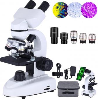
What Is Diaphragm In Microscope ?
The diaphragm in a microscope is a circular disk located beneath the stage that controls the amount of light that passes through the specimen. It is also known as the iris diaphragm because it functions similarly to the iris of the eye. By adjusting the diaphragm, the user can regulate the amount of light that enters the microscope, which can improve the clarity and contrast of the image. The diaphragm typically consists of a series of overlapping metal blades that can be opened or closed by rotating a lever or dial. When the diaphragm is fully open, the maximum amount of light is allowed to pass through the specimen. When it is closed, the amount of light is reduced, which can be useful when observing specimens that are particularly bright or when using high magnification lenses. Proper use of the diaphragm is an important aspect of microscope technique and can greatly enhance the quality of the images obtained.
1、 Structure and Location of the Diaphragm

The diaphragm in a microscope is a circular or semi-circular disc located beneath the stage. It is used to control the amount of light that passes through the specimen and into the objective lens. The diaphragm is made up of a series of adjustable blades that can be opened or closed to adjust the size of the aperture. By adjusting the size of the aperture, the amount of light that passes through the specimen can be controlled, which can help to improve the clarity and contrast of the image.
The diaphragm is an important component of the microscope, as it helps to regulate the amount of light that enters the objective lens. This is important because too much light can cause the image to appear washed out, while too little light can make it difficult to see the specimen clearly. By adjusting the diaphragm, the user can find the optimal balance of light and contrast to produce a clear and detailed image.
Recent advances in microscope technology have led to the development of new types of diaphragms, such as the iris diaphragm. This type of diaphragm uses a series of overlapping blades that can be adjusted to create a circular aperture of varying size. The iris diaphragm is particularly useful for high-magnification microscopy, as it allows for precise control over the amount of light that enters the objective lens.
In summary, the diaphragm in a microscope is a crucial component that helps to regulate the amount of light that enters the objective lens. By adjusting the size of the aperture, the user can find the optimal balance of light and contrast to produce a clear and detailed image. Recent advances in microscope technology have led to the development of new types of diaphragms, such as the iris diaphragm, which offer even greater control over the amount of light that enters the lens.
2、 Function of the Diaphragm

The diaphragm in a microscope is a circular disk located beneath the stage that controls the amount of light that passes through the specimen. It is also known as an iris diaphragm because it functions similarly to the iris of the human eye. The diaphragm has a series of adjustable blades that can be opened or closed to adjust the size of the aperture, which controls the amount of light that reaches the specimen.
The function of the diaphragm is to regulate the amount and quality of light that passes through the specimen. By adjusting the size of the aperture, the diaphragm can control the depth of field, contrast, and resolution of the image. A smaller aperture will increase the depth of field and improve resolution, while a larger aperture will decrease the depth of field and increase contrast.
Recent advancements in microscope technology have led to the development of automated diaphragms that can adjust the aperture based on the type of specimen being viewed. These automated diaphragms use algorithms to optimize the amount and quality of light that passes through the specimen, resulting in clearer and more detailed images.
In summary, the diaphragm in a microscope is an essential component that controls the amount and quality of light that passes through the specimen. Its function is to regulate the depth of field, contrast, and resolution of the image, and recent advancements in technology have led to the development of automated diaphragms that can optimize the image quality.
3、 Types of Diaphragms in Microscopes

What is diaphragm in microscope? The diaphragm in a microscope is a circular or semi-circular disc located beneath the stage. It is used to control the amount of light that passes through the specimen and into the objective lens. The diaphragm can be adjusted to change the size of the aperture, which in turn affects the amount of light that reaches the specimen. This is important because too much light can cause the specimen to appear washed out, while too little light can make it difficult to see.
There are several types of diaphragms in microscopes, including the iris diaphragm, the simple diaphragm, and the field diaphragm. The iris diaphragm is the most common type and is found in most modern microscopes. It consists of a series of overlapping metal blades that can be adjusted to change the size of the aperture. The simple diaphragm is a flat disc with a single hole that can be rotated to adjust the size of the aperture. The field diaphragm is a circular disc that is used to control the size of the field of view.
In recent years, there has been a growing interest in using computer-controlled diaphragms in microscopes. These diaphragms can be programmed to adjust the amount of light that passes through the specimen based on the specific needs of the user. This technology has the potential to greatly improve the accuracy and efficiency of microscopy, particularly in fields such as medical research and diagnostics.
4、 Adjusting the Diaphragm for Optimal Viewing

The diaphragm in a microscope is a circular or semi-circular disc located beneath the stage. It is used to control the amount of light that passes through the specimen and into the objective lens. By adjusting the diaphragm, the user can regulate the amount of light that enters the microscope, which can improve the clarity and contrast of the image.
To adjust the diaphragm for optimal viewing, the user should start by placing the specimen on the stage and selecting the appropriate objective lens. Next, the user should adjust the focus and brightness of the microscope to achieve a clear image. Once the image is in focus, the user can adjust the diaphragm to control the amount of light that enters the microscope.
If the image appears too bright or washed out, the user should close the diaphragm slightly to reduce the amount of light. Conversely, if the image appears too dark or lacks contrast, the user should open the diaphragm slightly to allow more light to enter the microscope.
It is important to note that the optimal diaphragm setting may vary depending on the type of specimen being viewed and the objective lens being used. Additionally, some modern microscopes may have automatic diaphragm adjustments that can optimize the image for the user.


