What Is Field Of View In Microscope ?
The field of view in a microscope refers to the area of the specimen that is visible through the eyepiece. It is the circular area that you see when you look through the microscope. The size of the field of view depends on the magnification of the microscope. At higher magnifications, the field of view becomes smaller, but the details of the specimen become more visible. The field of view is an important consideration when using a microscope because it affects the amount of detail that can be seen and the ease of locating specific structures within the specimen.
1、 Magnification
Field of view in a microscope refers to the area visible through the eyepiece or objective lens. It is the circular or rectangular area that is visible when looking through the microscope. The size of the field of view depends on the magnification of the microscope. As the magnification increases, the field of view decreases.
Magnification, on the other hand, refers to the ability of a microscope to enlarge an object. It is the ratio of the size of the image seen through the microscope to the actual size of the object. Magnification is an important factor in microscopy as it allows us to see details that are not visible to the naked eye.
The field of view and magnification are closely related. As the magnification increases, the field of view decreases. This is because at higher magnifications, the microscope is zooming in on a smaller area of the specimen, resulting in a smaller field of view. Conversely, at lower magnifications, the microscope is zooming out, resulting in a larger field of view.
It is important to note that the field of view and magnification are not the only factors that determine the quality of a microscope image. Other factors such as resolution, contrast, and depth of field also play a role in producing clear and detailed images.
In recent years, advances in technology have led to the development of new microscopy techniques that allow for higher magnifications and larger fields of view. For example, super-resolution microscopy techniques such as stimulated emission depletion (STED) microscopy and structured illumination microscopy (SIM) can achieve resolutions beyond the diffraction limit, allowing for the visualization of structures at the nanoscale level.
2、 Eyepiece
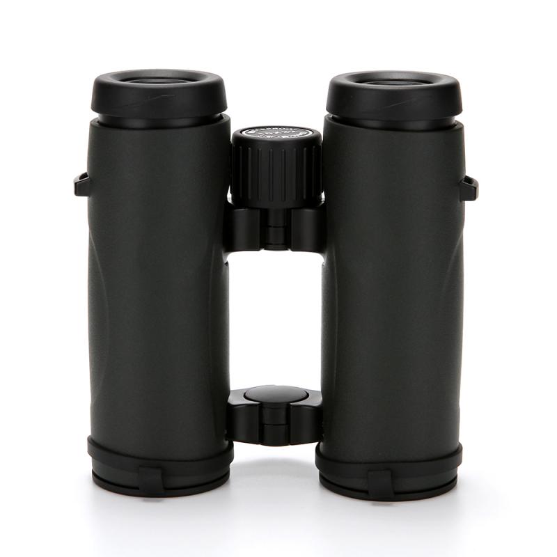
Field of view in a microscope refers to the area visible through the eyepiece when looking at a specimen. It is determined by the diameter of the eyepiece and the magnification of the objective lens. The higher the magnification, the smaller the field of view. The field of view is an important consideration when using a microscope because it affects the amount of detail that can be seen and the ease of locating specific structures within the specimen.
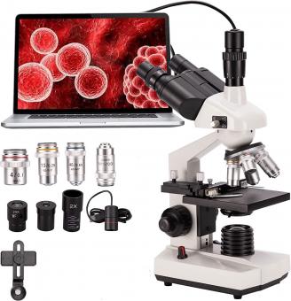
The eyepiece is an important component of the microscope that magnifies the image produced by the objective lens. It typically contains a set of lenses that further magnify the image and adjust the focus. The diameter of the eyepiece determines the size of the field of view, with larger eyepieces providing a wider field of view. The eyepiece also plays a role in determining the overall magnification of the microscope, as it is multiplied by the magnification of the objective lens to determine the total magnification.
Recent advances in microscope technology have led to the development of eyepieces with wider fields of view and higher magnification capabilities. These advancements have made it easier to observe and analyze specimens with greater detail and accuracy. Additionally, digital microscopes have become increasingly popular, allowing for the capture and analysis of images using computer software. These digital microscopes often have built-in cameras that provide a wider field of view and higher magnification than traditional eyepieces.
3、 Objective lens
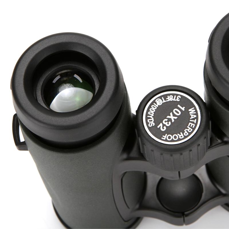
Objective lens is one of the most important components of a microscope. It is responsible for magnifying the specimen and producing a clear image. One of the key parameters associated with the objective lens is the field of view. The field of view in a microscope refers to the area of the specimen that is visible through the lens. It is determined by the diameter of the lens and the magnification power.
The field of view is an important consideration when using a microscope because it determines the amount of detail that can be observed. A larger field of view allows for a wider view of the specimen, but with less detail. Conversely, a smaller field of view provides a more detailed view of the specimen, but with a narrower view.
In recent years, there has been a growing interest in increasing the field of view in microscopes. This has been driven by the need to observe larger specimens, such as tissues and organs, and the desire to capture more information in a single image. To achieve this, new technologies have been developed, such as wide-field microscopy and panoramic imaging. These techniques use multiple lenses and advanced software algorithms to stitch together multiple images into a single, high-resolution image with a larger field of view.
In conclusion, the field of view is an important parameter associated with the objective lens in a microscope. It determines the amount of detail that can be observed and is a key consideration when using a microscope. With the development of new technologies, such as wide-field microscopy and panoramic imaging, the field of view in microscopes is increasing, allowing for the observation of larger specimens and the capture of more information in a single image.
4、 Numerical aperture
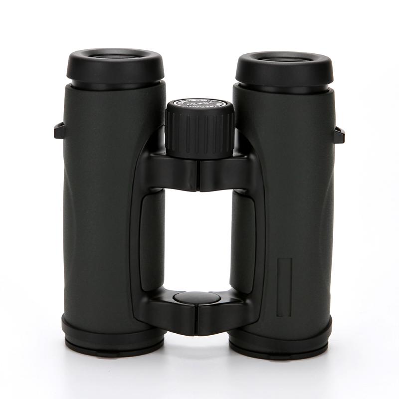
Field of view in a microscope refers to the area visible through the eyepiece or objective lens. It is the diameter of the circular image that is visible when looking through the microscope. The field of view is determined by the magnification of the objective lens and the size of the eyepiece. As the magnification increases, the field of view decreases.
Numerical aperture (NA) is a measure of the ability of an objective lens to gather and focus light. It is defined as the sine of the half-angle of the maximum cone of light that can enter the lens. The higher the numerical aperture, the greater the resolving power and the better the image quality. The numerical aperture is an important factor in determining the resolution of a microscope.
Recent advances in microscope technology have led to the development of high numerical aperture lenses, which have greatly improved the resolution of microscopes. These lenses allow for the visualization of structures that were previously too small to be seen with traditional microscopes. In addition, the use of fluorescence microscopy has enabled the visualization of specific molecules and structures within cells, further expanding the field of view in microscopy.
Overall, the field of view and numerical aperture are important factors in determining the quality and resolution of microscope images. Advances in technology continue to improve these factors, allowing for greater visualization of the microscopic world.


