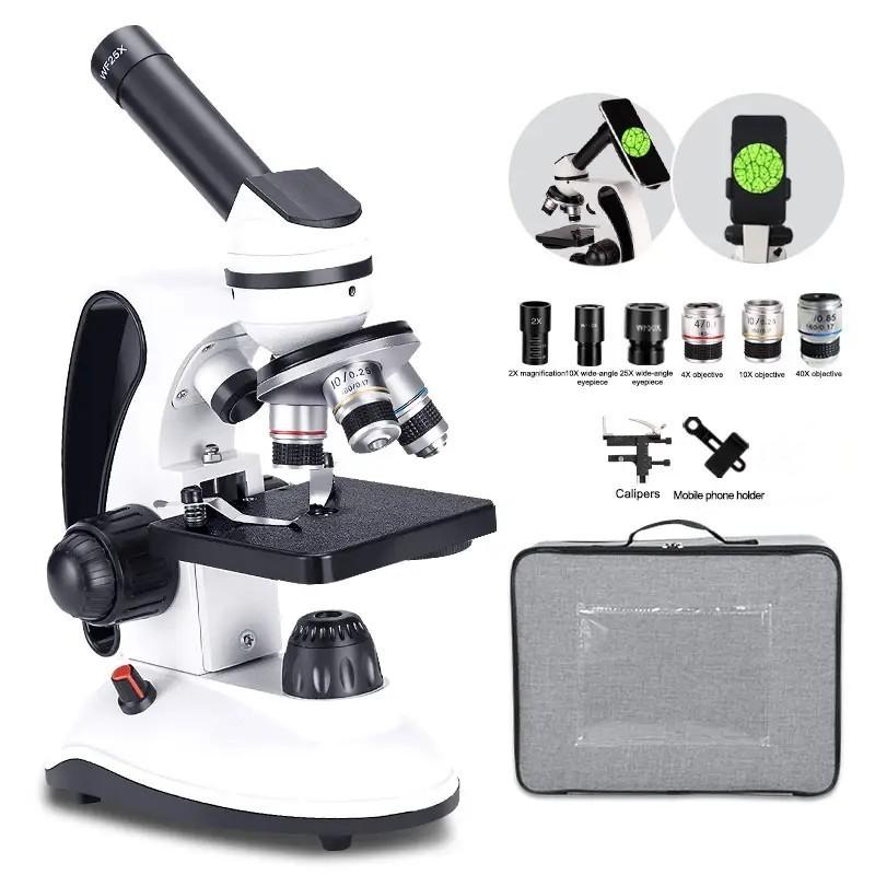
What Is Fluorescence Microscope ?
A fluorescence microscope is a type of microscope that uses fluorescence to visualize and study samples. It works by illuminating the sample with a specific wavelength of light, which causes certain molecules within the sample to emit light of a different color. This emitted light is then detected and used to create an image of the sample. Fluorescence microscopy is widely used in various fields of science, including biology, medicine, and materials science, as it allows researchers to study the structure and function of specific molecules within cells or tissues. It is particularly useful for visualizing fluorescently labeled molecules, such as fluorescent proteins or dyes, which can be used to highlight specific structures or molecules of interest.
1、 Principle of fluorescence microscopy
The fluorescence microscope is an advanced imaging tool used in various scientific fields, including biology, medicine, and materials science. It utilizes the principle of fluorescence to visualize and study specific molecules or structures within a sample.
Fluorescence microscopy works by exciting fluorescent molecules, called fluorophores, with a specific wavelength of light. When these molecules absorb the excitation light, they become temporarily energized and then emit light of a longer wavelength, known as fluorescence. This emitted light is then captured by the microscope's objective lens and detected by a camera or photomultiplier tube.
The principle of fluorescence microscopy allows for high-resolution imaging of specific molecules or structures within a sample. By labeling target molecules with fluorescent dyes or proteins, researchers can selectively visualize and study their distribution, localization, and dynamics within cells or tissues. This technique has revolutionized biological research, enabling the study of cellular processes, protein interactions, and disease mechanisms at the molecular level.
In recent years, advancements in fluorescence microscopy have further expanded its capabilities. Super-resolution techniques, such as stimulated emission depletion (STED) microscopy and single-molecule localization microscopy (SMLM), have pushed the limits of resolution beyond the diffraction barrier, allowing for the visualization of nanoscale structures and molecular interactions. Additionally, the development of genetically encoded fluorescent proteins, such as green fluorescent protein (GFP), has provided researchers with powerful tools for studying dynamic processes in living cells.
Overall, the fluorescence microscope is a versatile and indispensable tool in modern scientific research. Its ability to selectively visualize and study specific molecules or structures has greatly contributed to our understanding of biological processes and has the potential to drive further discoveries in various fields.

2、 Components of a fluorescence microscope
A fluorescence microscope is an advanced optical instrument used to visualize and study fluorescently labeled samples. It is designed to exploit the phenomenon of fluorescence, where certain molecules absorb light at a specific wavelength and emit light at a longer wavelength. This technique allows researchers to observe specific structures or molecules within a sample with high sensitivity and resolution.
The components of a fluorescence microscope include:
1. Light source: Typically, a high-intensity lamp or laser is used to provide the excitation light. The light is filtered to emit a specific wavelength that matches the excitation spectrum of the fluorophore being used.
2. Excitation filter: This filter selectively transmits the excitation wavelength, allowing only the desired light to reach the sample.
3. Dichroic mirror: Also known as a beamsplitter, this mirror reflects the excitation light towards the sample while allowing the emitted fluorescence to pass through.
4. Objective lens: This lens collects the emitted fluorescence from the sample and focuses it onto the detector.
5. Emission filter: This filter blocks the excitation light and allows only the emitted fluorescence to pass through to the detector.
6. Detector: Typically, a camera or a photomultiplier tube is used to capture the emitted fluorescence. These detectors convert the light into an electrical signal, which can be further processed and analyzed.
Recent advancements in fluorescence microscopy include the development of super-resolution techniques such as stimulated emission depletion (STED) microscopy and single-molecule localization microscopy (SMLM). These techniques allow researchers to surpass the diffraction limit of light, enabling the visualization of structures at the nanoscale level. Additionally, the integration of automated imaging systems and advanced image analysis algorithms has facilitated high-throughput imaging and quantitative analysis of fluorescence microscopy data.
In conclusion, a fluorescence microscope is a powerful tool that enables researchers to visualize and study fluorescently labeled samples with high sensitivity and resolution. Ongoing advancements in technology continue to enhance the capabilities of fluorescence microscopy, opening up new possibilities for biological and biomedical research.

3、 Fluorescent dyes and probes used in fluorescence microscopy
A fluorescence microscope is an advanced imaging tool that utilizes the phenomenon of fluorescence to visualize and study biological samples at a cellular and molecular level. It is a powerful technique widely used in various fields of research, including biology, medicine, and materials science.
Fluorescence microscopy involves the use of fluorescent dyes and probes that emit light of a specific wavelength when excited by a light source. These dyes and probes are designed to bind to specific molecules or structures within the sample, allowing researchers to selectively visualize and study them. The emitted light is then captured by the microscope and converted into an image, providing detailed information about the sample's composition, localization, and dynamics.
Fluorescent dyes and probes used in fluorescence microscopy have evolved significantly over the years, with continuous advancements in their design and performance. The latest developments focus on improving the brightness, photostability, and specificity of the dyes and probes. This enables researchers to achieve higher resolution and sensitivity in their imaging experiments.
Additionally, recent advancements in fluorescence microscopy techniques, such as super-resolution microscopy, have revolutionized the field by surpassing the diffraction limit of light. These techniques allow researchers to visualize structures and processes at an unprecedented level of detail, providing new insights into cellular and molecular biology.
Overall, fluorescence microscopy, with its ability to selectively label and visualize specific molecules, has become an indispensable tool in modern biological research. It continues to play a crucial role in advancing our understanding of complex biological systems and holds great potential for future discoveries.
4、 Applications of fluorescence microscopy in biological research
A fluorescence microscope is an advanced imaging tool that utilizes fluorescence to visualize and study biological samples. It is equipped with a light source that emits a specific wavelength of light, which excites fluorescent molecules within the sample. These molecules then emit light of a different wavelength, which is captured by the microscope's detectors and converted into an image.
Fluorescence microscopy has revolutionized biological research by enabling scientists to observe specific molecules or structures within cells and tissues with high sensitivity and resolution. It allows for the visualization of dynamic processes in living cells, such as protein interactions, cellular signaling, and gene expression. By labeling specific molecules with fluorescent tags, researchers can track their movement and behavior in real-time, providing valuable insights into cellular function and disease mechanisms.
The applications of fluorescence microscopy in biological research are vast. It has been instrumental in studying cellular processes like mitosis, cell division, and organelle dynamics. It has also been used to investigate the localization and function of proteins, DNA, RNA, and other biomolecules within cells. Additionally, fluorescence microscopy has been crucial in studying the interactions between pathogens and host cells, as well as in drug discovery and development.
In recent years, advancements in fluorescence microscopy techniques have further expanded its applications. Super-resolution microscopy techniques, such as stimulated emission depletion (STED) and structured illumination microscopy (SIM), have pushed the limits of resolution, allowing researchers to visualize structures at the nanoscale. Furthermore, the development of genetically encoded fluorescent proteins, such as green fluorescent protein (GFP), has enabled the visualization of specific proteins in living organisms without the need for external labeling.
Overall, fluorescence microscopy continues to be a powerful tool in biological research, providing valuable insights into the intricate workings of cells and tissues. Its versatility and ability to capture dynamic processes make it an indispensable technique in various fields, including cell biology, immunology, neuroscience, and cancer research.
