What Is Focusing In Microscope ?
Focusing in a microscope refers to the process of adjusting the position of the objective lens or the stage to obtain a clear and sharp image of the specimen being observed. This is achieved by moving the lens or the stage up or down until the focal point of the lens coincides with the plane of the specimen. Focusing is an essential step in microscopy as it allows the observer to see the details of the specimen at different magnifications. There are two types of focusing in a microscope: coarse and fine focusing. Coarse focusing involves moving the stage or the lens rapidly to bring the specimen into focus, while fine focusing involves making small adjustments to the lens or the stage to achieve a sharper image. The process of focusing may vary depending on the type of microscope being used, but the basic principles remain the same.
1、 Magnification
Focusing in a microscope refers to the process of adjusting the lenses to obtain a clear and sharp image of the specimen being observed. This is achieved by adjusting the distance between the objective lens and the specimen, as well as the distance between the eyepiece and the observer's eye.
Magnification, on the other hand, refers to the ability of a microscope to enlarge the image of the specimen being observed. This is achieved by using lenses with different focal lengths to increase the size of the image.
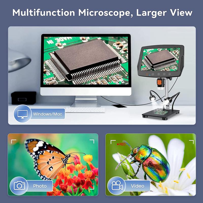
In recent years, there has been a growing interest in the use of digital microscopes, which use digital cameras to capture images of the specimen and display them on a computer screen. These microscopes often have built-in autofocus systems, which use algorithms to adjust the focus automatically, making it easier for users to obtain clear and sharp images.
Another recent development in microscopy is the use of super-resolution techniques, which allow for the observation of structures that are smaller than the diffraction limit of light. These techniques, such as stimulated emission depletion (STED) microscopy and structured illumination microscopy (SIM), use complex optical systems and advanced algorithms to achieve resolutions that were previously thought to be impossible.
Overall, focusing and magnification are essential aspects of microscopy, and recent advances in technology have made it easier than ever to obtain high-quality images of specimens at a range of scales.

2、 Resolution
What is focusing in microscope? Focusing in microscope refers to the process of adjusting the microscope's lenses to obtain a clear and sharp image of the specimen being observed. This is achieved by moving the objective lens closer or further away from the specimen until the image is in focus. The process of focusing is crucial in microscopy as it determines the clarity and quality of the image obtained.
Resolution, on the other hand, refers to the ability of a microscope to distinguish between two closely spaced objects. It is a measure of the microscope's ability to produce a clear and sharp image of the specimen being observed. The higher the resolution of a microscope, the better its ability to distinguish between two closely spaced objects.
In recent years, there have been significant advancements in microscopy technology, leading to the development of high-resolution microscopes such as the super-resolution microscope. These microscopes use advanced techniques such as stimulated emission depletion (STED) microscopy and structured illumination microscopy (SIM) to achieve resolutions beyond the diffraction limit of light. This has revolutionized the field of microscopy, allowing scientists to observe and study biological structures and processes at the molecular level.
In conclusion, focusing in microscope is the process of adjusting the lenses to obtain a clear and sharp image of the specimen being observed, while resolution refers to the ability of a microscope to distinguish between two closely spaced objects. With the development of high-resolution microscopes, scientists can now observe and study biological structures and processes at the molecular level, leading to new discoveries and advancements in the field of microscopy.

3、 Contrast
Focusing in a microscope refers to the process of adjusting the lenses to obtain a clear and sharp image of the specimen being observed. This is achieved by adjusting the distance between the objective lens and the specimen, as well as the distance between the eyepiece and the observer's eye.
Contrast, on the other hand, refers to the difference in brightness and color between the specimen and its background. This is important in microscopy as it allows for the visualization of fine details and structures within the specimen.
In recent years, there has been a growing interest in improving contrast in microscopy through the use of advanced techniques such as phase contrast, differential interference contrast, and fluorescence microscopy. These techniques allow for the visualization of structures that may not be visible with traditional brightfield microscopy.
Additionally, the development of digital imaging technology has allowed for the enhancement and manipulation of contrast in microscopy images. This has led to the creation of new tools and software that enable researchers to analyze and quantify the contrast of their images, providing valuable insights into the structure and function of biological specimens.
Overall, focusing and contrast are essential components of microscopy that continue to evolve and improve with advances in technology and techniques.
4、 Depth of Field
Depth of field is a term used in microscopy to describe the range of distance that is in focus at any given time. It is the distance between the nearest and farthest objects in a scene that appear acceptably sharp in an image. In other words, it is the area of the specimen that is in focus when viewed through the microscope.
Focusing in microscopy is the process of adjusting the microscope to achieve the desired depth of field. This is done by adjusting the focus knob to move the objective lens closer or further away from the specimen. The goal is to find the optimal position where the specimen is in focus and the depth of field is sufficient for the intended purpose.
The depth of field in microscopy is influenced by several factors, including the numerical aperture of the objective lens, the wavelength of light used, and the refractive index of the medium between the objective lens and the specimen. In recent years, advances in technology have led to the development of new microscopy techniques that allow for improved depth of field and resolution, such as confocal microscopy and super-resolution microscopy.
In conclusion, focusing in microscopy is the process of adjusting the microscope to achieve the desired depth of field, which is the range of distance that is in focus at any given time. The depth of field is influenced by several factors and can be improved with the use of advanced microscopy techniques.


