What Is High Magnification On A Microscope ?
High magnification on a microscope refers to the ability of the microscope to enlarge the image of the specimen being observed. It is a measure of how much larger the specimen appears compared to its actual size. High magnification allows for detailed examination of the specimen, revealing fine structures and features that may not be visible at lower magnifications. This is achieved by using objective lenses with high numerical aperture and short focal length, combined with appropriate eyepieces. The total magnification is determined by multiplying the magnification of the objective lens with that of the eyepiece. High magnification is particularly useful in fields such as biology, medicine, and materials science, where the observation of small structures is crucial for research and analysis.
1、 Resolution: Achieving fine detail and clarity in the magnified image.
High magnification on a microscope refers to the ability of the microscope to enlarge an object or specimen to a larger size. It allows scientists and researchers to observe and study minute details that are not visible to the naked eye. The magnification power of a microscope is determined by the combination of the objective lens and the eyepiece.
Resolution, on the other hand, refers to the ability of a microscope to achieve fine detail and clarity in the magnified image. It is a measure of how well the microscope can distinguish between two closely spaced objects. A higher resolution means that the microscope can provide a clearer and more detailed image.
Achieving high magnification and resolution is crucial in various scientific fields such as biology, medicine, and materials science. It allows scientists to study the intricate structures of cells, tissues, and microorganisms, leading to a better understanding of biological processes and disease mechanisms. In materials science, high magnification and resolution enable researchers to analyze the composition and structure of materials at the atomic and molecular level, aiding in the development of new materials with improved properties.
Advancements in technology have led to the development of more sophisticated microscopes with higher magnification and resolution capabilities. For instance, electron microscopes use a beam of electrons instead of light, allowing for much higher magnification and resolution. Scanning probe microscopes, such as atomic force microscopy, can achieve atomic-scale resolution by scanning a sharp probe over the surface of a sample.
In conclusion, high magnification on a microscope allows for the enlargement of objects or specimens, while resolution ensures fine detail and clarity in the magnified image. These capabilities are essential for scientific research and have been continuously improved with advancements in microscope technology.

2、 Numerical aperture: Determining the light-gathering ability of the microscope.
High magnification on a microscope refers to the ability of the microscope to enlarge the image of a specimen to a larger size. It allows scientists and researchers to observe minute details and structures that are not visible to the naked eye. High magnification is achieved by using a combination of lenses and optical systems that increase the size of the image.
One of the key factors that determine the high magnification capability of a microscope is the numerical aperture (NA). Numerical aperture is a measure of the light-gathering ability of the microscope. It is determined by the refractive index of the medium between the specimen and the objective lens, as well as the angle at which the light enters the lens. A higher numerical aperture allows more light to enter the lens, resulting in a higher resolution and better image quality at high magnifications.
In recent years, advancements in technology have led to the development of microscopes with even higher magnification capabilities. For example, super-resolution microscopy techniques such as stimulated emission depletion (STED) microscopy and structured illumination microscopy (SIM) have pushed the limits of resolution beyond the diffraction limit, allowing scientists to observe structures at the nanoscale.
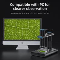
Furthermore, the integration of digital imaging and computer analysis has revolutionized microscopy. High-resolution cameras and sophisticated software enable researchers to capture and analyze images in real-time, enhancing the capabilities of high-magnification microscopy. This has opened up new avenues for research in various fields, including biology, medicine, materials science, and nanotechnology.
In conclusion, high magnification on a microscope is achieved through a combination of lenses and optical systems, with the numerical aperture playing a crucial role in determining the light-gathering ability of the microscope. Recent advancements in technology have further expanded the capabilities of high-magnification microscopy, allowing scientists to explore the microscopic world with unprecedented detail and resolution.

3、 Objective lenses: Selecting appropriate lenses for high magnification.
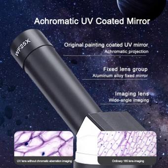
High magnification on a microscope refers to the ability of the microscope to enlarge the image of a specimen to a high degree. It is achieved by using objective lenses with a high numerical aperture (NA) and short focal length. These lenses are designed to capture more light and provide a greater level of detail and resolution in the magnified image.
Objective lenses are an essential component of a microscope and are responsible for gathering light from the specimen and forming an enlarged image. Selecting appropriate lenses for high magnification is crucial to achieve accurate and detailed observations. The choice of objective lenses depends on the specific requirements of the experiment or observation.
In recent years, there have been advancements in objective lens technology that have further improved high magnification capabilities. For example, the development of super-resolution microscopy techniques, such as stimulated emission depletion (STED) microscopy and structured illumination microscopy (SIM), has allowed researchers to achieve resolutions beyond the diffraction limit of light. These techniques involve using specialized objective lenses that can provide higher magnification and resolution.
Additionally, the use of advanced materials and coatings in objective lenses has improved their optical performance. For instance, the development of apochromatic lenses has reduced chromatic aberrations, resulting in sharper and more accurate images. Furthermore, the use of anti-reflective coatings on lens surfaces has minimized light loss and improved image contrast.
In conclusion, high magnification on a microscope is achieved by using objective lenses with a high numerical aperture and short focal length. Selecting appropriate lenses for high magnification is crucial for obtaining accurate and detailed observations. Recent advancements in objective lens technology, such as super-resolution microscopy techniques and improved lens materials and coatings, have further enhanced the capabilities of high magnification microscopy.

4、 Optical aberrations: Correcting distortions and imperfections in the magnified image.
High magnification on a microscope refers to the ability of the microscope to enlarge the image of a specimen to a greater extent. It allows for a more detailed examination of the specimen, revealing finer structures and features that may not be visible at lower magnifications. High magnification is achieved by using objective lenses with a higher numerical aperture and shorter focal length.
One of the challenges associated with high magnification is the occurrence of optical aberrations. These are distortions and imperfections in the magnified image that can affect the clarity and accuracy of the observed specimen. Common types of optical aberrations include spherical aberration, chromatic aberration, and coma.
To correct these aberrations, various techniques and technologies have been developed. For example, the use of aspherical lenses can help reduce spherical aberration, while the combination of multiple lenses with different refractive indices can minimize chromatic aberration. Additionally, advanced microscope designs, such as apochromatic objectives, are specifically engineered to correct for multiple types of aberrations simultaneously.
In recent years, there have been significant advancements in microscope technology aimed at improving high magnification imaging. For instance, the development of adaptive optics has allowed for real-time correction of optical aberrations, resulting in sharper and more accurate images. Furthermore, the integration of digital imaging and computational algorithms has enabled the enhancement and restoration of high-magnification images, compensating for any residual aberrations.
Overall, high magnification on a microscope provides researchers and scientists with the ability to explore the intricate details of specimens. While optical aberrations pose challenges, ongoing advancements in microscope technology continue to address and minimize these distortions, allowing for more precise and reliable observations at high magnifications.


There are no comments for this blog.