What Is Illumination In Microscope ?
Illumination in a microscope refers to the light source that is used to illuminate the specimen being observed. The light source can be located either above or below the specimen, depending on the type of microscope being used. In a compound microscope, for example, the light source is typically located below the specimen and is directed upward through the stage and into the objective lens. In a stereo microscope, on the other hand, the light source is typically located above the specimen and is directed downward onto the specimen.
The quality and intensity of the illumination can have a significant impact on the quality of the image produced by the microscope. Proper illumination is essential for producing clear, sharp images with good contrast and color. Different types of illumination techniques can be used depending on the type of specimen being observed and the specific requirements of the observation. For example, brightfield illumination is commonly used for observing stained specimens, while darkfield illumination is used for observing unstained specimens.
1、 Brightfield illumination
Brightfield illumination is a type of illumination used in microscopy that involves the use of a light source to illuminate the sample being observed. The light passes through the sample and is then collected by the objective lens, which magnifies the image and sends it to the eyepiece for observation. This type of illumination is commonly used in biological and medical research, as well as in industrial applications.
In brightfield illumination, the light source is typically located below the sample, and the sample is placed on a glass slide. The light passes through the sample and is absorbed or scattered by the various structures within the sample. The resulting image is a dark object on a bright background, which can be observed through the eyepiece.
One of the limitations of brightfield illumination is that it can be difficult to observe transparent or translucent samples, as they may not absorb or scatter enough light to produce a visible image. In recent years, new techniques such as phase contrast and differential interference contrast (DIC) microscopy have been developed to address this limitation and improve the visualization of transparent samples.
Overall, brightfield illumination remains a widely used and important technique in microscopy, particularly in the fields of biology and medicine. Its simplicity and versatility make it a valuable tool for a wide range of applications, from basic research to clinical diagnosis.
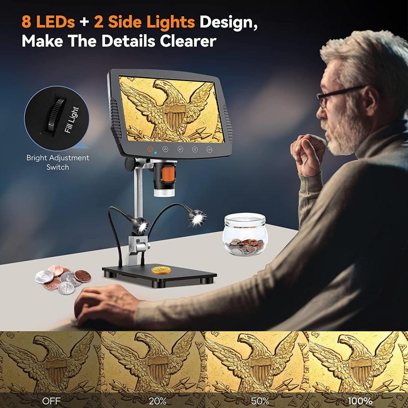
2、 Darkfield illumination
Darkfield illumination is a technique used in microscopy to visualize specimens that are difficult to see under brightfield illumination. In darkfield illumination, the specimen is illuminated with a cone of light that is directed at an angle to the optical axis of the microscope. This causes the light to scatter off the specimen, creating a bright image against a dark background.
The advantage of darkfield illumination is that it can reveal details that are not visible under brightfield illumination. For example, it can be used to visualize bacteria, which are often too small to be seen under brightfield illumination. It can also be used to visualize transparent or translucent specimens, such as living cells or small organisms.
One of the latest developments in darkfield illumination is the use of digital microscopy. Digital microscopy allows for the capture of high-resolution images and videos of specimens under darkfield illumination. This has led to new insights into the behavior and structure of microorganisms, as well as the development of new diagnostic tools for diseases.
In conclusion, darkfield illumination is a powerful technique in microscopy that allows for the visualization of specimens that are difficult to see under brightfield illumination. With the advent of digital microscopy, it is now possible to capture high-resolution images and videos of specimens under darkfield illumination, leading to new insights and applications in the field of microbiology.

3、 Phase contrast illumination
Phase contrast illumination is a technique used in microscopy to enhance the contrast of transparent and colorless specimens. It works by transforming the phase differences of light waves passing through the specimen into differences in brightness, which can be visualized by the observer. This technique is particularly useful for observing living cells and tissues, as it allows for the visualization of internal structures and movements that would otherwise be difficult to see.
In phase contrast illumination, a special condenser lens is used to create a phase shift in the light passing through the specimen. This phase shift is then converted into a difference in brightness by a phase plate located in the objective lens. The resulting image appears as a bright specimen on a dark background, which enhances the contrast and visibility of the specimen.
Recent advancements in phase contrast microscopy have led to the development of new techniques, such as quantitative phase imaging and digital holography. These techniques use phase contrast illumination to measure the refractive index of the specimen, which can provide information about its physical properties and composition. This has applications in fields such as cell biology, materials science, and nanotechnology.
Overall, phase contrast illumination is a powerful tool in microscopy that allows for the visualization of transparent and colorless specimens. Its continued development and application in new fields will undoubtedly lead to further discoveries and advancements in science and technology.

4、 Differential interference contrast (DIC) illumination
Differential interference contrast (DIC) illumination is a type of illumination used in microscopy to enhance the contrast of transparent and semi-transparent specimens. DIC illumination works by splitting a beam of light into two beams that pass through the specimen at slightly different angles. The two beams are then recombined, creating an interference pattern that highlights the edges and boundaries of the specimen.
DIC illumination is particularly useful for imaging living cells and tissues, as it can reveal details that are difficult to see with other types of illumination. It can also be used to visualize structures such as organelles and membranes, which are often too small or too transparent to be seen with conventional brightfield microscopy.
Recent advances in DIC illumination technology have led to the development of new techniques such as structured illumination microscopy (SIM) and super-resolution structured illumination microscopy (SR-SIM). These techniques use patterned illumination to further enhance the resolution and contrast of DIC images, allowing researchers to see even finer details in their specimens.
Overall, DIC illumination is a powerful tool for microscopy that has revolutionized our ability to study the structure and function of living cells and tissues. As technology continues to advance, it is likely that we will see even more sophisticated forms of DIC illumination that will further expand our understanding of the microscopic world.


