What Is Phase Contrast Microscope Used For ?
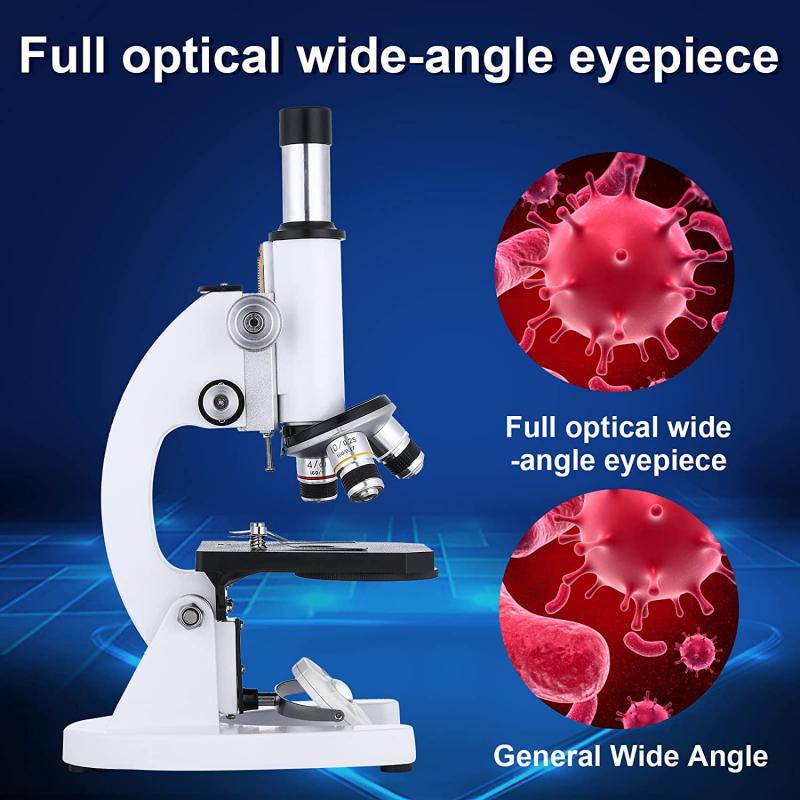
Phase contrast microscope is a type of light microscope that is used to visualize transparent and unstained specimens, such as living cells, microorganisms, and tissues. It enhances the contrast of these specimens by exploiting the differences in their refractive indices, which affect the speed and direction of light passing through them.
The phase contrast microscope is particularly useful in biological and medical research, as it allows scientists to observe the internal structures and movements of living cells and microorganisms without killing or staining them. It is commonly used in fields such as microbiology, cell biology, developmental biology, and pathology.
In addition to its research applications, the phase contrast microscope is also used in clinical settings for the diagnosis of various diseases, such as malaria, tuberculosis, and cancer. It can also be used in industrial and environmental monitoring to detect and analyze microorganisms and other small particles in water, food, and other samples.
1、 Optical microscopy technique
Phase contrast microscopy is an optical microscopy technique that is used to enhance the contrast of transparent and colorless specimens. It is widely used in biological research to observe living cells and tissues without the need for staining or fixing. The technique works by converting the phase differences in light passing through a specimen into differences in brightness, which can be visualized using a phase contrast microscope.
The phase contrast microscope is particularly useful for observing cells and tissues that are difficult to see with other microscopy techniques. For example, it can be used to observe the movement of cells, such as the beating of cilia or the flow of cytoplasm. It can also be used to observe the structure of cells, such as the shape and size of organelles.
In addition to its use in biological research, phase contrast microscopy is also used in other fields, such as materials science and engineering. It can be used to observe the structure of materials, such as polymers and ceramics, and to study the behavior of fluids, such as the flow of blood in microfluidic devices.
The latest point of view on phase contrast microscopy is that it is a powerful tool for studying biological systems at the cellular and subcellular level. It allows researchers to observe living cells and tissues in real-time, without the need for invasive techniques such as staining or fixing. As such, it is an important tool for advancing our understanding of biological processes and for developing new treatments for diseases.
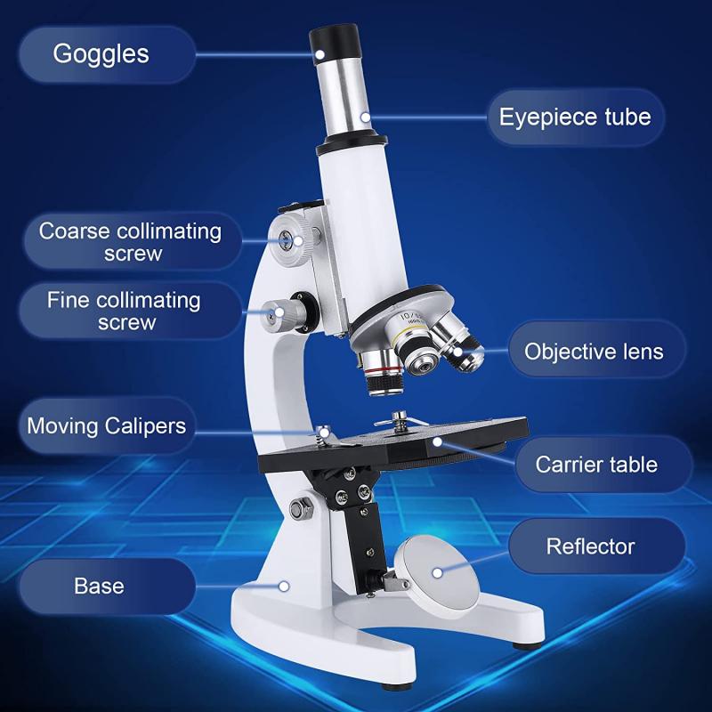
2、 Enhances contrast of transparent specimens
The phase contrast microscope is a type of light microscope that is used to enhance the contrast of transparent specimens. It is particularly useful for observing living cells and tissues, as it allows researchers to view the internal structures of these specimens without the need for staining or other invasive techniques.
The phase contrast microscope works by exploiting the differences in the refractive index of different parts of a specimen. As light passes through the specimen, it is slowed down by the denser parts of the specimen, causing a phase shift in the light waves. This phase shift is then converted into differences in brightness and contrast by the microscope, allowing the internal structures of the specimen to be visualized.
In addition to its use in biological research, the phase contrast microscope is also used in a variety of other fields, including materials science, geology, and engineering. For example, it can be used to study the microstructure of metals and alloys, or to observe the behavior of fluids and gases in microfluidic devices.
Overall, the phase contrast microscope is a powerful tool for observing and analyzing transparent specimens, and its versatility and ease of use make it a valuable asset in a wide range of scientific disciplines.
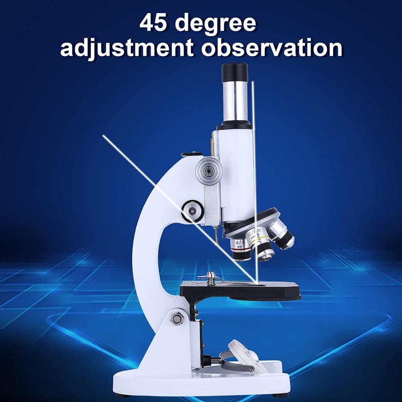
3、 Utilizes phase shifts of light passing through specimen
The phase contrast microscope is a type of light microscope that utilizes phase shifts of light passing through a specimen to produce high-contrast images of transparent and unstained samples. This technique is particularly useful for observing living cells and tissues, as it allows for the visualization of structures that would otherwise be invisible under normal brightfield microscopy.
The phase contrast microscope works by converting the phase differences between the light waves passing through the specimen and those passing through the surrounding medium into differences in brightness or contrast. This creates an image that highlights the subtle variations in refractive index within the specimen, such as the edges of cells or the internal structures of organelles.
In addition to its use in biological research, the phase contrast microscope has also found applications in materials science, where it can be used to study the microstructure of materials such as polymers, ceramics, and metals. It has also been used in the study of microfluidics, where it can be used to visualize the flow of fluids through small channels and devices.
Overall, the phase contrast microscope is a powerful tool for studying the structure and behavior of living cells and tissues, as well as a wide range of other materials and systems. Its ability to produce high-contrast images of transparent samples has made it an essential tool in many fields of research, and its continued development and refinement are likely to lead to even more exciting applications in the future.
4、 Allows visualization of internal structures
The phase contrast microscope is a type of light microscope that allows for the visualization of internal structures of cells and tissues that would otherwise be difficult to see with traditional brightfield microscopy. This is achieved by exploiting the differences in refractive index between different parts of the specimen, which causes variations in the phase of the light passing through it.
The phase contrast microscope is commonly used in biological research and medical diagnostics to study living cells and tissues. It is particularly useful for observing cells in culture, as it allows researchers to monitor cell growth and behavior over time without damaging the cells. It is also used in the study of microorganisms, such as bacteria and fungi, as well as in the examination of blood and other bodily fluids.
In recent years, the phase contrast microscope has been used in a variety of new applications, including the study of nanomaterials and the development of new materials for use in electronics and other industries. It has also been used in the field of neuroscience to study the structure and function of neurons and other cells in the brain.
Overall, the phase contrast microscope is a powerful tool for visualizing internal structures and processes in a wide range of biological and materials science applications. Its versatility and ease of use make it an essential tool for researchers and clinicians alike.
