What Is The Diaphragm On A Microscope ?
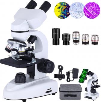
The diaphragm on a microscope is a circular or semi-circular disk located beneath the stage of the microscope. It is used to control the amount of light that passes through the specimen being observed. The diaphragm has several small holes of varying sizes that can be adjusted to regulate the amount of light that enters the microscope. By adjusting the diaphragm, the user can control the contrast and brightness of the image being viewed. This is particularly important when observing specimens that are transparent or have low contrast. The diaphragm is an essential component of a microscope and is used in conjunction with other components such as the objective lenses and eyepiece to produce a clear and detailed image of the specimen.
1、 Optical Microscopy
What is the diaphragm on a microscope? The diaphragm is a component of the microscope that controls the amount of light that passes through the specimen. It is located beneath the stage and consists of a series of adjustable apertures that can be opened or closed to regulate the amount of light that reaches the specimen. By adjusting the diaphragm, the user can control the contrast and brightness of the image, as well as reduce glare and improve resolution.
In optical microscopy, the diaphragm is an essential component that helps to optimize the quality of the image. It is particularly important when working with specimens that are highly reflective or have low contrast, as it can help to reduce unwanted reflections and improve the visibility of the specimen. Additionally, the diaphragm can be used to adjust the depth of field, which is the range of distances that are in focus at any given time.
Recent advances in microscopy technology have led to the development of new types of diaphragms that offer even greater control over the light that passes through the specimen. For example, some microscopes now feature electronically controlled diaphragms that can be adjusted with a high degree of precision. Other microscopes use specialized diaphragms that are designed to work with specific types of specimens, such as those that are highly reflective or have complex structures.
Overall, the diaphragm is a critical component of the microscope that plays a key role in optimizing the quality of the image. As microscopy technology continues to evolve, it is likely that new types of diaphragms will be developed that offer even greater control and precision.

2、 Microscope Components
The diaphragm on a microscope is a component that controls the amount of light that passes through the specimen being observed. It is located beneath the stage and consists of a series of adjustable apertures that can be opened or closed to regulate the amount of light that enters the microscope. The diaphragm is an essential component of the microscope as it helps to improve the clarity and contrast of the image being viewed.
The diaphragm works by controlling the angle of the light that enters the microscope. When the aperture is opened wider, more light enters the microscope, which can be useful when observing specimens that are particularly dark or opaque. Conversely, when the aperture is closed down, less light enters the microscope, which can be useful when observing specimens that are particularly bright or transparent.
In recent years, there has been a trend towards using digital microscopes, which use cameras and computer software to capture and analyze images. While these microscopes do not have physical diaphragms, they often have software-based controls that allow users to adjust the amount of light that enters the microscope. This allows for greater flexibility and precision when observing specimens, and can be particularly useful when working with delicate or sensitive samples.

3、 Diaphragm Function
What is the diaphragm on a microscope? The diaphragm is a small, circular or square-shaped disc located beneath the stage of a microscope. It is used to control the amount of light that passes through the specimen being viewed. By adjusting the diaphragm, the user can regulate the amount of light that enters the microscope, which can help to improve the clarity and contrast of the image.
The diaphragm works by adjusting the size of the aperture through which light passes. When the diaphragm is opened wider, more light is allowed to enter the microscope, which can be useful when viewing specimens that are particularly dark or opaque. Conversely, when the diaphragm is closed down, less light is allowed to enter the microscope, which can be useful when viewing specimens that are particularly bright or transparent.
In addition to controlling the amount of light that enters the microscope, the diaphragm can also be used to adjust the depth of field. By adjusting the diaphragm, the user can change the amount of light that is focused on the specimen, which can help to bring different parts of the specimen into focus.
Overall, the diaphragm is an important component of a microscope that helps to control the amount of light that enters the instrument. By adjusting the diaphragm, users can improve the clarity and contrast of the image, as well as adjust the depth of field.

4、 Types of Diaphragms
What is the diaphragm on a microscope? The diaphragm on a microscope is a circular or semi-circular disc located beneath the stage of the microscope. It is used to control the amount of light that passes through the specimen being viewed. The diaphragm can be adjusted to change the size of the aperture, which in turn controls the amount of light that enters the microscope. This is important because too much light can cause the specimen to appear washed out, while too little light can make it difficult to see the details of the specimen.
There are several types of diaphragms that can be used on a microscope. The most common type is the iris diaphragm, which is a circular disc with adjustable blades that can be opened or closed to control the amount of light that enters the microscope. Another type of diaphragm is the simple diaphragm, which is a fixed disc with a small hole in the center that can be rotated to adjust the amount of light that enters the microscope.
In recent years, there has been a growing interest in using digital microscopes, which use cameras and computer software to capture and analyze images of specimens. These microscopes often have built-in diaphragms that can be controlled using software, allowing for precise adjustments to the amount of light that enters the microscope. This can be particularly useful for researchers who need to capture high-quality images of specimens for analysis and publication.


There are no comments for this blog.