What Is The Function Of Microscope ?
The function of a microscope is to magnify small objects or organisms that are not visible to the naked eye. It allows for detailed examination and study of the structure, composition, and behavior of these objects. Microscopes are widely used in various scientific fields, such as biology, medicine, chemistry, and materials science, to observe and analyze microscopic samples. They work by using lenses or a combination of lenses to focus light or electrons, enabling the user to see the specimen with enhanced clarity and resolution. Microscopes can be classified into different types, including optical microscopes, electron microscopes, and scanning probe microscopes, each with its own specific applications and capabilities.
1、 Magnification: Enlarging objects to see fine details.
The function of a microscope is to magnify objects and enable us to see fine details that are not visible to the naked eye. Magnification is the primary function of a microscope, as it allows us to enlarge objects and observe them in greater detail. By using lenses and other optical components, microscopes can increase the size of an object, making it easier to study its structure and characteristics.
Microscopes have been used for centuries in various scientific fields, such as biology, medicine, and materials science. They have played a crucial role in advancing our understanding of the natural world and have contributed to numerous scientific discoveries. With the ability to magnify objects, microscopes have allowed scientists to observe and study cells, microorganisms, and other tiny structures that are essential to our understanding of life and the natural world.
In recent years, advancements in technology have led to the development of more sophisticated microscopes. For example, electron microscopes use a beam of electrons instead of light to magnify objects, allowing for even higher levels of magnification and resolution. This has opened up new possibilities in fields such as nanotechnology and materials science, where the ability to observe and manipulate objects at the atomic and molecular level is crucial.
Furthermore, the integration of digital imaging and computer technology has revolutionized microscopy. Digital microscopes can capture high-resolution images and videos, allowing for easier documentation and sharing of findings. Additionally, advanced software and image analysis techniques enable researchers to extract quantitative data from microscope images, enhancing the accuracy and efficiency of their studies.
In conclusion, the primary function of a microscope is to magnify objects and enable us to see fine details. However, with advancements in technology, microscopes have evolved to offer higher levels of magnification, resolution, and imaging capabilities. These advancements have expanded the applications of microscopy and continue to contribute to scientific discoveries and advancements in various fields.

2、 Resolution: Ability to distinguish between closely spaced objects.
The function of a microscope is to magnify and enhance the visibility of objects that are too small to be seen by the naked eye. One crucial aspect of a microscope's functionality is its ability to provide resolution, which refers to its capacity to distinguish between closely spaced objects. Resolution is a fundamental characteristic of microscopes as it determines the level of detail that can be observed and analyzed.
The resolution of a microscope is determined by its optical system, specifically the quality of its lenses and the wavelength of light used. The higher the resolution, the more detail can be seen in the specimen. Microscopes with high resolution are essential in various scientific fields, such as biology, medicine, and materials science, where the examination of minute structures is crucial.
In recent years, there have been significant advancements in microscope technology, leading to improved resolution capabilities. For instance, the development of super-resolution microscopy techniques has revolutionized the field by surpassing the traditional diffraction limit of light. Techniques like stimulated emission depletion (STED) microscopy and structured illumination microscopy (SIM) have allowed scientists to visualize structures at the nanoscale level, providing unprecedented insights into cellular processes and molecular interactions.
Furthermore, advancements in electron microscopy have also contributed to enhancing resolution. Transmission electron microscopy (TEM) and scanning electron microscopy (SEM) have become invaluable tools for studying the ultrastructure of cells and materials, enabling researchers to observe objects with resolutions down to the atomic level.
In conclusion, the function of a microscope is to magnify and enhance the visibility of small objects, with resolution being a critical aspect of its functionality. The ability to distinguish between closely spaced objects allows scientists to observe and analyze intricate details, leading to advancements in various scientific disciplines. With the continuous development of microscopy techniques, the resolution capabilities of microscopes have significantly improved, enabling researchers to explore the microscopic world with unprecedented clarity and precision.

3、 Illumination: Providing light to enhance visibility of the specimen.
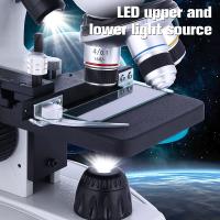
The function of a microscope is to magnify small objects or specimens, allowing for detailed observation and analysis. One crucial aspect of a microscope is its illumination system, which provides light to enhance the visibility of the specimen. Illumination plays a vital role in microscopy as it determines the quality and clarity of the image produced.
The primary purpose of illumination is to ensure that the specimen is adequately lit, allowing for better visibility and contrast. By illuminating the specimen, the microscope enables the observer to see fine details that would otherwise be difficult to discern. This is particularly important when studying transparent or translucent specimens, as proper illumination helps to reveal their internal structures and features.
In addition to enhancing visibility, illumination also affects the resolution and image quality. The type and intensity of light used can influence the level of detail that can be observed. Different illumination techniques, such as brightfield, darkfield, phase contrast, and fluorescence, offer unique advantages for specific types of specimens and research purposes.
With advancements in technology, the function of illumination in microscopy has evolved. Modern microscopes often incorporate advanced lighting systems, such as LED or fiber optic illumination, which provide brighter and more uniform lighting. These systems offer benefits such as longer lifespan, energy efficiency, and reduced heat generation compared to traditional light sources like halogen bulbs.
Furthermore, the latest developments in microscopy, such as confocal microscopy and super-resolution microscopy, have revolutionized the field by providing even higher resolution and improved imaging capabilities. These techniques often utilize specialized illumination methods, such as laser illumination, to achieve exceptional image quality and enable researchers to explore the microscopic world in unprecedented detail.
In conclusion, the function of illumination in microscopy is to provide light that enhances the visibility of the specimen, allowing for detailed observation and analysis. Illumination not only improves visibility but also influences the resolution and image quality. With advancements in technology, the role of illumination in microscopy has expanded, offering brighter and more uniform lighting, as well as enabling cutting-edge techniques for high-resolution imaging.

4、 Focus: Adjusting the sharpness and clarity of the image.
The function of a microscope is to magnify small objects or organisms that are not visible to the naked eye, allowing scientists and researchers to study them in detail. One crucial aspect of using a microscope is the ability to focus and adjust the sharpness and clarity of the image. This is achieved by manipulating the focus knobs on the microscope.
The focus adjustment is essential because it determines the level of detail that can be observed. By adjusting the focus, scientists can bring the object or organism into clear view, enabling them to examine its structure, morphology, and other characteristics. This process is particularly important when studying cells, microorganisms, or tiny particles, as even the slightest adjustment can reveal new information.
In recent years, advancements in microscope technology have led to the development of more sophisticated focusing mechanisms. For example, some microscopes now incorporate autofocus systems that automatically adjust the focus based on the object being observed. This feature has greatly improved the efficiency and accuracy of microscopic observations, especially in fields such as medical diagnostics and biological research.
Additionally, digital microscopes have emerged, which allow for real-time imaging and analysis. These microscopes often have software that enables users to adjust the focus digitally, providing even greater control and precision. This digital focus adjustment has proven to be particularly useful in fields like materials science and nanotechnology, where precise measurements and analysis are crucial.
In conclusion, the function of a microscope is to magnify small objects and organisms, and focusing is a fundamental aspect of this process. The ability to adjust the sharpness and clarity of the image allows scientists and researchers to observe and study microscopic structures in detail, leading to advancements in various scientific disciplines.


