What Microscope Can See Bacteria ?
A compound microscope is the most commonly used microscope to see bacteria. It uses two or more lenses to magnify the image of the specimen. The lenses bend the light passing through the specimen, which makes the image appear larger. Compound microscopes can magnify up to 1000 times, which is enough to see most bacteria. However, some bacteria are too small to be seen with a compound microscope, and an electron microscope is needed to visualize them. Electron microscopes use a beam of electrons instead of light to magnify the image, which allows for much higher magnification and resolution.
1、 Compound Microscopes
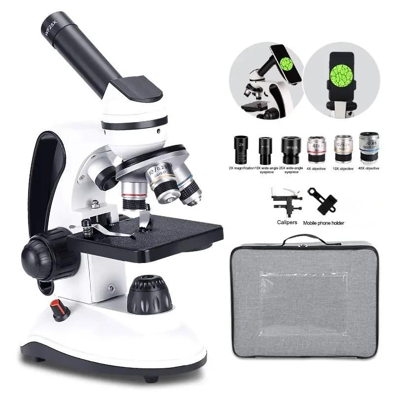
Compound microscopes are the most commonly used type of microscope for observing bacteria. These microscopes use two or more lenses to magnify the image of the specimen, allowing for a detailed view of the bacteria. The lenses in a compound microscope are arranged in a series, with the objective lens closest to the specimen and the eyepiece lens closest to the observer's eye.
With the use of compound microscopes, scientists have been able to study bacteria in great detail, including their morphology, structure, and behavior. They have also been able to identify different types of bacteria and study their interactions with other organisms.
In recent years, advances in technology have led to the development of more sophisticated microscopes, such as electron microscopes and super-resolution microscopes. These microscopes have allowed scientists to study bacteria at an even higher level of detail, revealing the intricate structures and processes that occur within bacterial cells.
Despite these advances, compound microscopes remain an essential tool for studying bacteria, particularly in microbiology and medical research. They are relatively inexpensive, easy to use, and provide a clear and detailed view of bacterial specimens. As such, they are likely to remain a staple of scientific research for years to come.
2、 Darkfield Microscopes
What microscope can see bacteria? Darkfield microscopes are one type of microscope that can visualize bacteria. These microscopes use a special condenser that illuminates the specimen with oblique light, causing the bacteria to appear as bright objects against a dark background. This technique is particularly useful for observing live, unstained bacteria, as it allows for better contrast and resolution than other types of microscopy.
In recent years, advances in technology have led to the development of new microscopy techniques that can visualize bacteria in even greater detail. For example, super-resolution microscopy techniques such as structured illumination microscopy (SIM) and stimulated emission depletion (STED) microscopy can achieve resolutions of less than 100 nanometers, allowing for the visualization of individual molecules within bacterial cells.
Another emerging technique is single-molecule localization microscopy (SMLM), which uses fluorescent probes to tag individual molecules within bacterial cells. By analyzing the positions of these fluorescent probes, researchers can reconstruct high-resolution images of bacterial structures with resolutions of less than 20 nanometers.
Overall, while darkfield microscopy remains a valuable tool for visualizing bacteria, advances in microscopy technology are continually pushing the boundaries of what we can see and understand about these tiny organisms.
3、 Phase Contrast Microscopes
Phase Contrast Microscopes are the type of microscope that can see bacteria. These microscopes use a special optical technique that enhances the contrast of transparent and colorless specimens, such as bacteria, making them visible under the microscope. This technique works by converting the differences in refractive index of the specimen into differences in brightness, which allows the bacteria to be seen clearly.
Phase Contrast Microscopes have been used for many years to study bacteria and other microorganisms. They are widely used in microbiology, medical research, and other fields where the study of microorganisms is important. However, with the advancement of technology, newer and more advanced microscopes have been developed that can also see bacteria.
One such microscope is the Fluorescence Microscope, which uses fluorescent dyes to label specific structures or molecules within the bacteria, making them visible under the microscope. Another type of microscope is the Electron Microscope, which uses a beam of electrons to create an image of the bacteria at a much higher resolution than traditional light microscopes.
Despite the availability of newer and more advanced microscopes, Phase Contrast Microscopes remain an important tool in the study of bacteria. They are relatively simple to use, require minimal sample preparation, and are widely available in research laboratories and educational institutions.

4、 Fluorescence Microscopes
Fluorescence microscopes are one of the types of microscopes that can see bacteria. These microscopes use fluorescent dyes or proteins that bind to specific structures within the bacteria, causing them to emit light when illuminated with a specific wavelength of light. This allows for the visualization of bacteria that would otherwise be invisible under a traditional brightfield microscope.
Fluorescence microscopy has revolutionized the study of bacteria, allowing researchers to study the behavior and interactions of individual bacterial cells in real-time. This has led to a better understanding of bacterial physiology, pathogenesis, and antibiotic resistance.
In recent years, advances in fluorescence microscopy have further expanded its capabilities. Super-resolution fluorescence microscopy techniques, such as stimulated emission depletion (STED) microscopy and structured illumination microscopy (SIM), have allowed for the visualization of bacterial structures at a resolution beyond the diffraction limit of light. This has enabled researchers to study the intricate details of bacterial structures, such as the bacterial cell wall and flagella, with unprecedented clarity.
Overall, fluorescence microscopy has become an essential tool in the study of bacteria, providing researchers with a powerful tool to investigate the complex world of bacterial cells.


