What Microscope Can See Mitochondria ?
A transmission electron microscope (TEM) is capable of visualizing mitochondria.
1、 Light Microscopy: Limited resolution, mitochondria appear as small dots.
Light microscopy is a widely used technique in biological research and can provide valuable insights into cellular structures, including mitochondria. However, due to its limited resolution, mitochondria appear as small dots under a light microscope. This is because the resolution of light microscopy is limited by the wavelength of visible light, which is around 400-700 nanometers.
Mitochondria are small organelles, typically measuring 1-10 micrometers in length, and their intricate internal structures cannot be resolved in detail using a light microscope. While the overall shape and distribution of mitochondria can be observed, finer details such as the inner mitochondrial membrane, cristae, and matrix are not visible with this technique.
To overcome the limitations of light microscopy, researchers often employ other advanced imaging techniques such as electron microscopy or super-resolution microscopy. Electron microscopy uses a beam of electrons instead of light, allowing for much higher resolution imaging. With electron microscopy, the internal structures of mitochondria can be visualized in great detail, providing insights into their morphology and organization.
Super-resolution microscopy techniques, such as stimulated emission depletion (STED) microscopy or structured illumination microscopy (SIM), can also surpass the diffraction limit of light microscopy. These techniques use various approaches to achieve resolutions beyond the limits of conventional light microscopy, enabling the visualization of finer details within mitochondria.
In recent years, advancements in microscopy techniques have allowed for even more detailed imaging of mitochondria. For example, correlative light and electron microscopy (CLEM) combines the advantages of both light and electron microscopy, allowing researchers to observe mitochondria at different levels of resolution and gain a more comprehensive understanding of their structure and function.
In conclusion, while light microscopy can provide a general overview of mitochondria, it has limited resolution and cannot reveal the finer details of their internal structures. To visualize mitochondria in greater detail, researchers often turn to electron microscopy or super-resolution microscopy techniques. These advanced imaging methods have significantly contributed to our understanding of mitochondria and their role in cellular processes.
2、 Electron Microscopy: High resolution, reveals detailed mitochondrial structure.
Electron Microscopy: High resolution, reveals detailed mitochondrial structure.
Electron microscopy is a powerful tool that can be used to visualize mitochondria with high resolution and reveal their detailed structure. Mitochondria are small organelles found in most eukaryotic cells and are responsible for producing energy in the form of ATP. They have a complex internal structure, including an outer membrane, an inner membrane with folds called cristae, and a matrix containing enzymes and DNA.
Electron microscopy uses a beam of electrons instead of light to image samples. This allows for much higher resolution than traditional light microscopy, as electrons have much shorter wavelengths than visible light. With electron microscopy, scientists can visualize mitochondria at the nanometer scale, revealing intricate details of their structure.
In recent years, advancements in electron microscopy techniques have further improved our understanding of mitochondria. Cryo-electron microscopy, for example, allows samples to be imaged at extremely low temperatures, preserving their native structure. This technique has provided unprecedented insights into the organization and dynamics of mitochondrial membranes and proteins.
Furthermore, electron tomography, a three-dimensional imaging technique, has allowed researchers to reconstruct the entire mitochondrial structure in great detail. This has led to discoveries about the organization of cristae and their role in mitochondrial function.
Overall, electron microscopy is an essential tool for studying mitochondria and has greatly contributed to our understanding of their structure and function. With ongoing advancements in electron microscopy techniques, we can expect even more detailed insights into the complex world of mitochondria in the future.
3、 Confocal Microscopy: Provides 3D visualization of mitochondria within cells.
Confocal Microscopy: Provides 3D visualization of mitochondria within cells.
Confocal microscopy is a powerful imaging technique that allows for the visualization of mitochondria within cells in three dimensions. This technique has revolutionized the field of cell biology by providing detailed information about the structure and function of mitochondria.
Mitochondria are small organelles found in most eukaryotic cells that play a crucial role in energy production. They are responsible for generating adenosine triphosphate (ATP), the main energy currency of the cell. Understanding the structure and function of mitochondria is essential for studying various cellular processes, including metabolism, cell signaling, and cell death.
Confocal microscopy uses a laser beam to scan the sample and create high-resolution images. It works by illuminating a single plane within the sample, while blocking out-of-focus light from above and below that plane. This allows for the collection of sharp, clear images of mitochondria within cells.
The 3D visualization provided by confocal microscopy enables researchers to study the spatial distribution and organization of mitochondria within cells. It allows for the examination of mitochondrial dynamics, such as fission and fusion events, as well as their interactions with other cellular structures.
In recent years, advancements in confocal microscopy have further enhanced its capabilities for studying mitochondria. For example, the development of super-resolution techniques, such as stimulated emission depletion (STED) microscopy and structured illumination microscopy (SIM), has allowed for even higher resolution imaging of mitochondria. These techniques overcome the diffraction limit of light, enabling the visualization of finer details within the mitochondria.
Overall, confocal microscopy is a valuable tool for studying mitochondria within cells. Its ability to provide 3D visualization and high-resolution imaging has greatly contributed to our understanding of the structure and function of these essential organelles.
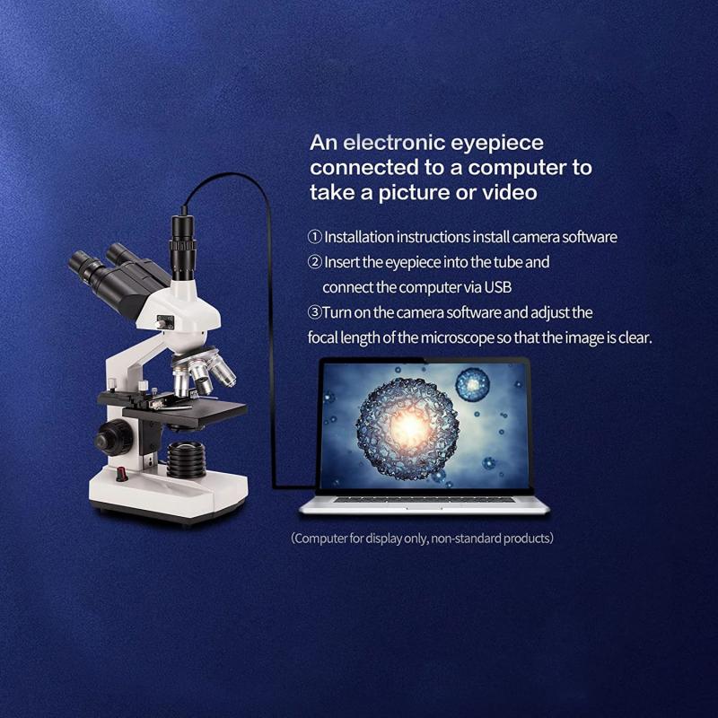
4、 Super-resolution Microscopy: Overcomes diffraction limit, enhances mitochondrial imaging.
Super-resolution microscopy is a powerful tool that can overcome the diffraction limit of conventional light microscopy, allowing for enhanced imaging of cellular structures such as mitochondria. Mitochondria are small organelles found in most eukaryotic cells and are responsible for producing energy in the form of ATP. They play a crucial role in cellular metabolism and are involved in various cellular processes.
Super-resolution microscopy techniques, such as stimulated emission depletion (STED) microscopy, structured illumination microscopy (SIM), and single-molecule localization microscopy (SMLM), have revolutionized the field of cell biology by providing unprecedented resolution and detail. These techniques utilize different principles to achieve super-resolution imaging, but they all share the ability to visualize structures that were previously beyond the diffraction limit of light microscopy.
By using super-resolution microscopy, researchers can obtain high-resolution images of mitochondria, revealing their intricate structure and dynamics. This allows for a better understanding of mitochondrial function and their involvement in various cellular processes, including cell signaling, apoptosis, and metabolism.
The latest advancements in super-resolution microscopy have further improved the imaging of mitochondria. For example, recent studies have combined super-resolution microscopy with fluorescent labeling techniques to visualize specific mitochondrial proteins or markers, providing insights into their localization and interactions within the organelle. Additionally, the development of new fluorescent probes and dyes specifically targeting mitochondria has enhanced the specificity and sensitivity of mitochondrial imaging.
In conclusion, super-resolution microscopy techniques have revolutionized the field of cell biology by enabling enhanced imaging of cellular structures, including mitochondria. These techniques have overcome the diffraction limit of conventional light microscopy, providing unprecedented resolution and detail. The latest advancements in super-resolution microscopy have further improved the imaging of mitochondria, allowing for a better understanding of their structure and function in cellular processes.
