What Microscope Is Used For Gram Staining ?
The microscope commonly used for Gram staining is a compound light microscope.
1、 Light microscope
The microscope used for gram staining is typically a light microscope. Gram staining is a common technique used in microbiology to differentiate bacteria into two major groups: gram-positive and gram-negative. This staining method involves a series of steps that include applying crystal violet, iodine, alcohol, and safranin to the bacterial sample. The gram-positive bacteria retain the crystal violet stain, appearing purple under the microscope, while the gram-negative bacteria lose the stain and appear pink or red.
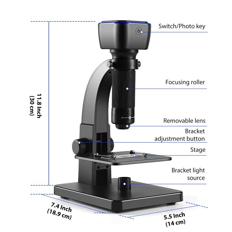
A light microscope is the most commonly used microscope for gram staining because it allows for the visualization of the stained bacteria at a cellular level. This type of microscope uses visible light to illuminate the sample and magnify the image. It typically consists of a light source, lenses, and an eyepiece or camera for viewing the specimen.
While there have been advancements in microscopy technology, such as electron microscopes and confocal microscopes, these are not commonly used for gram staining. Electron microscopes use a beam of electrons instead of light to magnify the sample, providing higher resolution images. However, they require specialized sample preparation techniques and are not suitable for live or stained bacterial samples.
Confocal microscopes, on the other hand, use laser light and a pinhole aperture to eliminate out-of-focus light, resulting in sharper images. While confocal microscopy can provide detailed images of stained bacteria, it is not typically used for gram staining due to its higher cost and complexity.
In conclusion, the light microscope remains the most practical and widely used microscope for gram staining. It allows for the visualization of stained bacteria at a cellular level, providing valuable information for microbiological research and diagnostics.
2、 Compound microscope
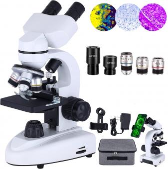
The compound microscope is commonly used for gram staining. Gram staining is a technique used in microbiology to differentiate bacteria into two major groups: Gram-positive and Gram-negative. This staining method involves the use of crystal violet dye, iodine, alcohol, and safranin. The compound microscope is essential for observing the stained bacteria and determining their Gram status.
A compound microscope consists of two or more lenses that work together to magnify the image of the specimen. It provides high magnification and resolution, allowing for detailed examination of the stained bacteria. The stained bacteria can be observed under different magnifications, ranging from low power (10x) to high power (100x or higher), depending on the specific requirements of the analysis.
In recent years, there have been advancements in microscopy techniques, such as the development of fluorescence microscopy and confocal microscopy. These techniques offer enhanced visualization and imaging capabilities, allowing for more detailed analysis of bacterial structures and interactions. However, when it comes to gram staining, the compound microscope remains the most commonly used tool due to its simplicity, affordability, and effectiveness in observing the stained bacteria.
It is worth noting that with the advancement of technology, there may be alternative microscopy techniques that can be used for gram staining. However, the compound microscope continues to be the standard tool in most microbiology laboratories for this purpose.

3、 Brightfield microscope
The microscope used for gram staining is typically a Brightfield microscope. This type of microscope is commonly used in microbiology laboratories for various staining techniques, including gram staining.
A Brightfield microscope is a basic and widely used microscope that allows for the observation of stained specimens. It uses a bright light source located beneath the specimen, which passes through the specimen and into the objective lens. The objective lens then magnifies the image, which is further magnified by the eyepiece lens for observation.
Gram staining is a differential staining technique used to classify bacteria into two main groups: gram-positive and gram-negative. It involves the application of a series of dyes to the bacterial cells, which allows for the differentiation of their cell wall structures. The gram-positive bacteria retain the crystal violet dye and appear purple under the microscope, while the gram-negative bacteria do not retain the dye and appear pink or red.
The Brightfield microscope is ideal for gram staining because it provides good contrast between the stained bacteria and the background. The bright light source allows for clear visualization of the stained cells, making it easier to differentiate between gram-positive and gram-negative bacteria. Additionally, the magnification capabilities of the Brightfield microscope enable the observation of individual bacterial cells and their cell wall structures.
It is worth noting that advancements in microscopy technology have led to the development of more specialized microscopes, such as fluorescence microscopes and confocal microscopes, which can also be used for gram staining. These advanced microscopes offer enhanced visualization and imaging capabilities, allowing for more detailed analysis of bacterial cells. However, the Brightfield microscope remains the most commonly used microscope for gram staining due to its simplicity, affordability, and effectiveness in routine laboratory settings.

4、 Darkfield microscope
The Darkfield microscope is not typically used for Gram staining. Gram staining is a technique used to differentiate bacteria into two major groups - Gram-positive and Gram-negative - based on their cell wall composition. This staining method involves the use of crystal violet dye, iodine, alcohol, and safranin to visualize the bacteria under a light microscope.
The Darkfield microscope, on the other hand, is a specialized type of microscope that is used to observe unstained, transparent specimens that do not absorb or scatter light. It is particularly useful for visualizing live, motile microorganisms such as bacteria, as it enhances the contrast between the specimen and the background.
While the Darkfield microscope is not directly used for Gram staining, it can be employed in conjunction with other techniques to study bacteria. For example, after performing Gram staining, the stained bacteria can be observed under a Darkfield microscope to study their morphology, motility, and arrangement. This can provide additional information about the bacteria and aid in their identification.
It is important to note that microscopy techniques are constantly evolving, and new advancements are being made to improve the visualization and analysis of microorganisms. Therefore, it is possible that in the future, new microscopy techniques may be developed that combine the benefits of both Gram staining and Darkfield microscopy, allowing for more comprehensive and detailed analysis of bacteria.


















There are no comments for this blog.