What Microscope To See Bacteria ?
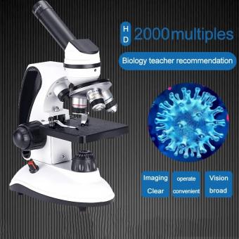
A compound light microscope is commonly used to see bacteria. This type of microscope uses visible light and a series of lenses to magnify the specimen. Bacteria are typically too small to be seen with the naked eye, so a microscope is necessary to observe them. A compound light microscope can magnify the specimen up to 1000 times, allowing for detailed observation of the bacteria's shape, size, and structure. In addition, specialized staining techniques can be used to make the bacteria more visible under the microscope. Other types of microscopes, such as electron microscopes, can also be used to observe bacteria, but they require more specialized equipment and expertise.
1、 Compound Microscopes
The best microscope to see bacteria is a compound microscope. This type of microscope uses two or more lenses to magnify the specimen, allowing for a clear and detailed view of the bacteria. Compound microscopes are commonly used in microbiology labs and are essential for studying bacteria and other microorganisms.
In recent years, there have been advancements in microscopy technology, such as the development of super-resolution microscopy. These new techniques allow for even higher magnification and resolution, which can provide more detailed information about the structure and behavior of bacteria. However, these techniques are still relatively new and may not be widely available or practical for routine use in most labs.
Overall, the compound microscope remains the most commonly used and practical tool for studying bacteria. It is relatively affordable, easy to use, and provides sufficient magnification and resolution for most applications. As technology continues to advance, we may see new microscopy techniques that offer even greater insights into the world of bacteria and other microorganisms.

2、 Darkfield Microscopes
What microscope to see bacteria? Darkfield microscopes are one of the best options for observing bacteria. These microscopes use a special condenser that blocks the direct light from the microscope's light source, causing the bacteria to appear as bright, glowing objects against a dark background. This technique allows for the visualization of bacteria that may be difficult to see with other types of microscopes.
In addition to their ability to visualize bacteria, darkfield microscopes have other advantages. They are relatively easy to use and require minimal sample preparation, making them a popular choice for microbiology labs. They are also useful for observing live bacteria, as the darkfield technique does not require staining or fixing the sample.
However, it is important to note that darkfield microscopes have limitations. They are not suitable for observing bacteria at high magnifications, as the dark background can become too bright and obscure the bacteria. Additionally, darkfield microscopes are not capable of providing detailed information about the internal structure of bacteria.
Overall, darkfield microscopes are a valuable tool for observing bacteria and are widely used in microbiology research. However, they should be used in conjunction with other microscopy techniques to obtain a complete understanding of bacterial structure and function.
3、 Phase Contrast Microscopes
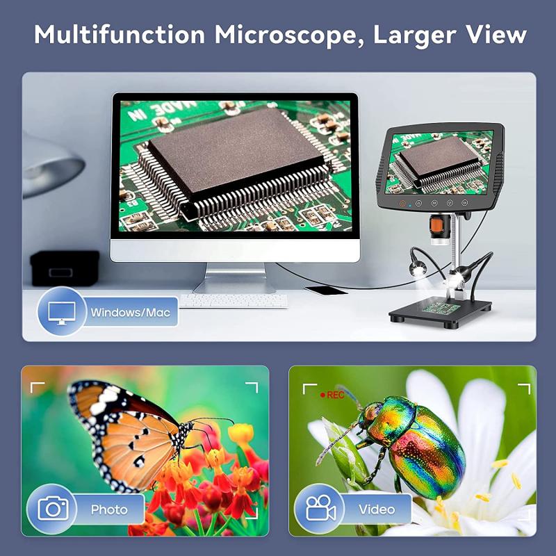
What microscope to see bacteria? The answer is Phase Contrast Microscopes. These microscopes are specifically designed to view transparent specimens, such as bacteria, that are difficult to see with traditional brightfield microscopes. Phase contrast microscopy allows for the visualization of live, unstained specimens without the need for special preparation techniques.
Phase contrast microscopes work by converting differences in refractive index into differences in brightness, creating contrast in the image. This allows for the visualization of structures within the bacteria, such as the cell wall, cytoplasm, and flagella. Additionally, phase contrast microscopy can be used to observe bacterial motility, which is important for understanding bacterial behavior and pathogenesis.
In recent years, advancements in technology have led to the development of digital phase contrast microscopy, which allows for the capture of high-resolution images and videos of live bacteria. This technology has been particularly useful in the study of bacterial biofilms, which are complex communities of bacteria that are difficult to observe with traditional microscopy techniques.
Overall, phase contrast microscopy is an essential tool for the study of bacteria and has contributed greatly to our understanding of bacterial structure, behavior, and pathogenesis. With continued advancements in technology, we can expect to see even more exciting developments in the field of bacterial microscopy in the years to come.
4、 Fluorescence Microscopes
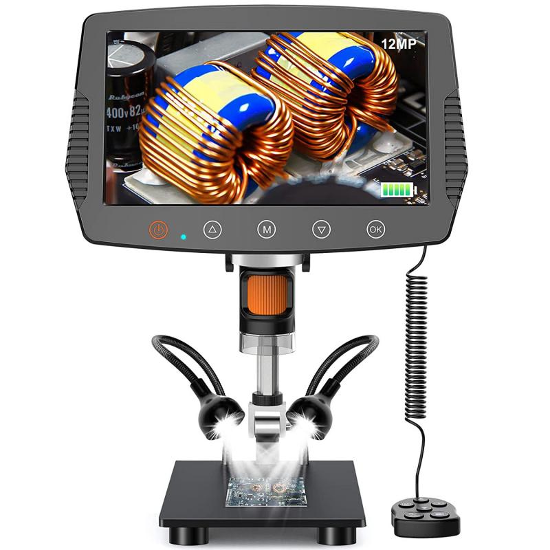
What microscope to see bacteria? Fluorescence microscopes are one of the best options for visualizing bacteria. These microscopes use fluorescent dyes or proteins that bind specifically to bacterial structures, allowing them to be easily seen under the microscope. Fluorescence microscopy has revolutionized the study of bacteria, allowing researchers to observe bacterial behavior and interactions in real-time.
One of the latest advancements in fluorescence microscopy is super-resolution microscopy, which allows for even higher resolution imaging of bacterial structures. This technique uses specialized fluorescent dyes and advanced imaging algorithms to achieve resolutions beyond the diffraction limit of traditional microscopy. This has allowed researchers to visualize bacterial structures at the nanoscale level, providing new insights into bacterial behavior and function.
Another recent development in fluorescence microscopy is the use of live-cell imaging, which allows researchers to observe bacterial behavior in real-time. This technique uses specialized microscopes and imaging software to capture images of living bacteria as they grow and interact with their environment. This has led to new discoveries about bacterial behavior and has the potential to improve our understanding of bacterial infections and how to treat them.
In conclusion, fluorescence microscopy, including super-resolution microscopy and live-cell imaging, is an excellent tool for visualizing bacteria. These techniques have revolutionized the study of bacteria and continue to provide new insights into bacterial behavior and function.


