What Size Microscope To See Bacteria ?
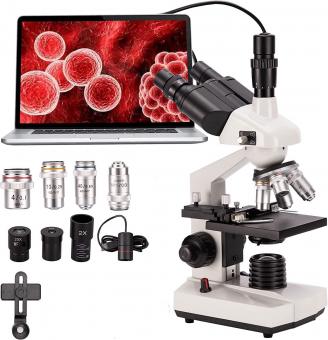
To see bacteria under a microscope, a high-power compound microscope is typically used. A microscope with a magnification of at least 400x is recommended to observe bacteria effectively. This level of magnification allows for the visualization of individual bacterial cells and their structures. Additionally, an oil immersion lens may be required for higher magnification, typically 1000x, to observe smaller bacteria or finer details. It is important to note that the size of bacteria can vary significantly, with some species being as small as 0.2 micrometers in diameter. Therefore, a microscope with adjustable magnification and good resolution is essential for accurate observation and identification of bacteria.
1、 Light Microscopy: Resolving Bacteria with Compound Light Microscopes
To see bacteria using a microscope, a compound light microscope is typically used. This type of microscope uses visible light to illuminate the specimen and magnify it for observation. The resolution of a microscope determines its ability to distinguish between two closely spaced objects, and this is crucial when trying to visualize bacteria.
The size of bacteria can vary, with most bacteria falling within the range of 0.2 to 2 micrometers in diameter. To observe bacteria with a compound light microscope, a resolution of at least 0.2 micrometers is necessary. This means that the microscope should have a magnification power of at least 1000x, as the maximum resolution of a light microscope is around 0.2 micrometers.
It is important to note that while a compound light microscope can visualize bacteria, it may not provide the level of detail required for certain applications. For more detailed examination, higher magnification and resolution microscopes such as electron microscopes are used. Electron microscopes use a beam of electrons instead of light, allowing for much higher magnification and resolution.
In recent years, advancements in microscopy techniques have allowed for even better visualization of bacteria. Super-resolution microscopy techniques, such as stimulated emission depletion (STED) microscopy and structured illumination microscopy (SIM), have pushed the limits of resolution beyond the diffraction limit of light. These techniques have enabled scientists to observe bacteria and other cellular structures with unprecedented detail.
In conclusion, a compound light microscope with a resolution of at least 0.2 micrometers and a magnification power of 1000x is suitable for visualizing bacteria. However, for more detailed examination, advanced microscopy techniques such as electron microscopy or super-resolution microscopy may be necessary.

2、 Electron Microscopy: Visualizing Bacteria with Electron Microscopes
To visualize bacteria, electron microscopy is the most suitable technique due to its high resolution capabilities. Electron microscopes use a beam of electrons instead of light to magnify the sample, allowing for much greater detail to be observed. This is crucial when studying bacteria, as they are typically smaller than the limit of resolution of light microscopes.
There are two types of electron microscopes commonly used to visualize bacteria: transmission electron microscopes (TEM) and scanning electron microscopes (SEM). TEMs are used to study the internal structure of bacteria, while SEMs provide a detailed view of the surface.
In terms of size, bacteria are generally in the range of 0.2 to 10 micrometers. To visualize bacteria using electron microscopy, a resolution of at least 0.1 micrometers is required. Both TEMs and SEMs are capable of achieving this level of resolution.
However, it is important to note that electron microscopy requires specialized equipment and expertise. Sample preparation for electron microscopy is more complex compared to light microscopy, involving fixation, dehydration, and staining. Additionally, electron microscopes are expensive and require a controlled environment to operate effectively.
In recent years, advancements in electron microscopy techniques have further improved the visualization of bacteria. Cryo-electron microscopy (cryo-EM) allows samples to be imaged at cryogenic temperatures, preserving their native structure. This technique has revolutionized the field of structural biology, enabling the visualization of bacteria and other biological specimens in unprecedented detail.
In conclusion, to visualize bacteria, electron microscopy is the most suitable technique. Both TEMs and SEMs can be used, with a resolution of at least 0.1 micrometers required. Recent advancements, such as cryo-EM, have further enhanced the visualization of bacteria, providing valuable insights into their structure and function.

3、 Optical Resolution: Achieving Sufficient Magnification for Bacterial Observation
To observe bacteria using a microscope, it is important to consider the optical resolution and magnification capabilities of the instrument. The size of bacteria typically ranges from 0.2 to 10 micrometers, making them relatively small and requiring a microscope with sufficient magnification.
The optical resolution of a microscope refers to its ability to distinguish two closely spaced objects as separate entities. In the case of bacteria, a microscope with high optical resolution is necessary to visualize the fine details and structures of these microorganisms. Achieving sufficient magnification is crucial to observe bacteria effectively.
In general, a compound light microscope is commonly used to observe bacteria. These microscopes use visible light and a series of lenses to magnify the specimen. With a standard compound light microscope, a magnification of around 1000x is typically sufficient to observe bacteria. This level of magnification allows for the visualization of individual bacterial cells and their structures.
However, it is important to note that the size and shape of bacteria can vary significantly. Some bacteria are larger and easier to observe, while others are smaller and require higher magnification. In recent years, advancements in microscopy techniques have allowed for even higher magnification capabilities. For example, the development of electron microscopy has enabled scientists to observe bacteria at much higher magnifications, up to several hundred thousand times.
In conclusion, to observe bacteria using a microscope, a compound light microscope with a magnification of around 1000x is generally sufficient. However, the size and characteristics of the bacteria being observed may require higher magnification capabilities. Advancements in microscopy techniques continue to push the boundaries of what is possible, allowing for even more detailed observations of bacteria.

4、 Numerical Aperture: Determining the Optimal Microscope Lens for Bacteria
Determining the optimal microscope lens for observing bacteria involves considering various factors, including the numerical aperture (NA) of the lens. The numerical aperture is a measure of the lens's ability to gather light and resolve fine details. A higher numerical aperture allows for better resolution and the ability to observe smaller objects, such as bacteria.
To see bacteria, a microscope with a relatively high numerical aperture is required. Bacteria are typically in the range of 0.2 to 2 micrometers in size, with some exceptions. To visualize these small organisms, a lens with a numerical aperture of at least 0.4 is recommended. This allows for sufficient resolution to observe the individual bacteria and their structures.
However, it is important to note that the optimal numerical aperture may vary depending on the specific bacteria being observed. Some bacteria have smaller sizes or unique characteristics that may require a higher numerical aperture for proper visualization. Additionally, advancements in microscopy techniques and technology may influence the optimal numerical aperture for observing bacteria.
Recent developments in microscopy, such as super-resolution techniques, have pushed the limits of resolution beyond the traditional diffraction limit. These techniques, such as stimulated emission depletion (STED) microscopy and structured illumination microscopy (SIM), allow for the visualization of bacteria and other cellular structures at a higher resolution than previously possible. These advancements have expanded our understanding of bacterial structures and interactions.
In conclusion, to observe bacteria using a microscope, a lens with a numerical aperture of at least 0.4 is recommended. However, the optimal numerical aperture may vary depending on the specific bacteria and advancements in microscopy techniques.


