What To Look At Through A Microscope ?
Through a microscope, one can observe a wide range of specimens and structures that are not visible to the naked eye. Some common things to look at through a microscope include cells, bacteria, fungi, viruses, tissues, and organs. Other objects that can be examined under a microscope include minerals, crystals, fibers, and small organisms such as protozoa and algae. Additionally, one can observe the structure of various materials such as metals, plastics, and ceramics. The use of a microscope is not limited to scientific research and can also be used in fields such as education, medicine, and industry.
1、 Magnification and Resolution
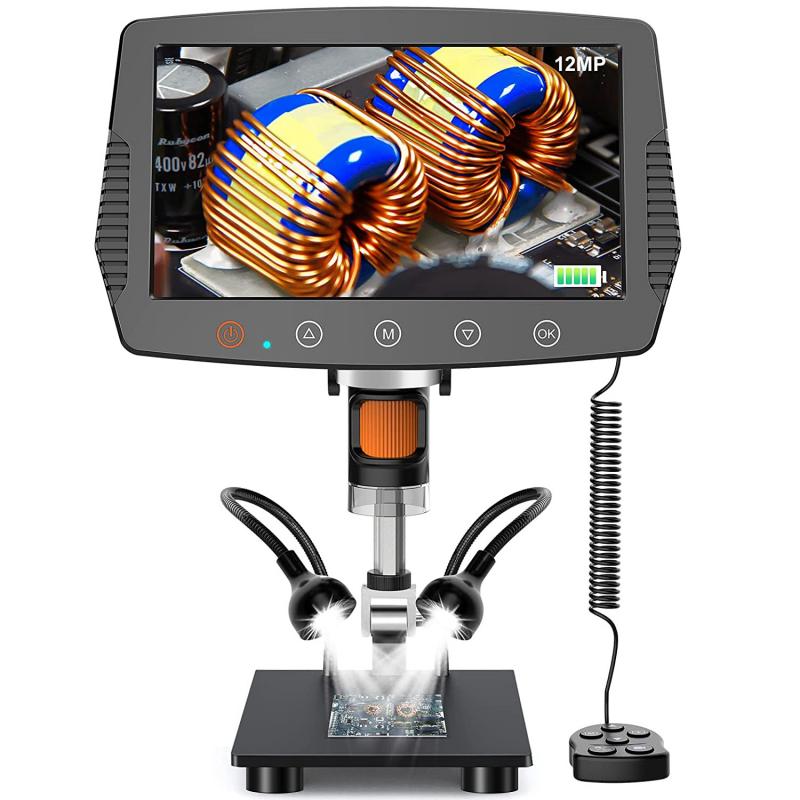
When looking through a microscope, there are several things to consider, but two of the most important are magnification and resolution. Magnification refers to how much larger an object appears under the microscope compared to its actual size, while resolution refers to the ability to distinguish between two closely spaced objects.
When choosing a microscope, it is important to consider the level of magnification needed for the specific application. For example, a low magnification microscope may be suitable for observing larger specimens such as insects or plant cells, while a higher magnification microscope may be necessary for observing smaller structures such as bacteria or viruses.
Resolution is also an important factor to consider when choosing a microscope. The ability to distinguish between two closely spaced objects is determined by the numerical aperture of the microscope lens and the wavelength of the light used to illuminate the specimen. In recent years, advances in technology have led to the development of super-resolution microscopy techniques, which allow for even higher levels of resolution and the ability to observe structures at the nanoscale.
In addition to magnification and resolution, other factors to consider when choosing a microscope include the type of illumination used, the type of specimen being observed, and the level of detail required. Ultimately, the choice of microscope will depend on the specific application and the level of detail required for the observation.
2、 Sample Preparation Techniques

Sample preparation techniques are crucial in microscopy as they determine the quality of the images obtained. When looking at samples through a microscope, it is important to consider the type of sample and the intended observation. Here are some things to look at through a microscope:
1. Cells and tissues: When observing cells and tissues, it is important to use fixation techniques to preserve the structure and prevent degradation. Staining techniques can also be used to enhance contrast and highlight specific structures.
2. Microorganisms: Microorganisms can be observed using wet mounts, where a drop of the sample is placed on a slide and covered with a coverslip. Staining techniques can also be used to enhance contrast and highlight specific structures.
3. Crystals: Crystals can be observed using polarized light microscopy, which allows for the visualization of crystal structure and orientation.
4. Nanoparticles: Nanoparticles can be observed using electron microscopy, which provides high-resolution images of the particles.
5. Biological molecules: Biological molecules such as proteins and DNA can be observed using techniques such as X-ray crystallography and cryo-electron microscopy, which provide detailed information about the structure and function of these molecules.
In recent years, there has been a growing interest in the use of super-resolution microscopy techniques, which allow for the visualization of structures at a resolution beyond the diffraction limit of light. These techniques have revolutionized the field of microscopy and have enabled researchers to study biological structures and processes in unprecedented detail.
3、 Types of Microscopes
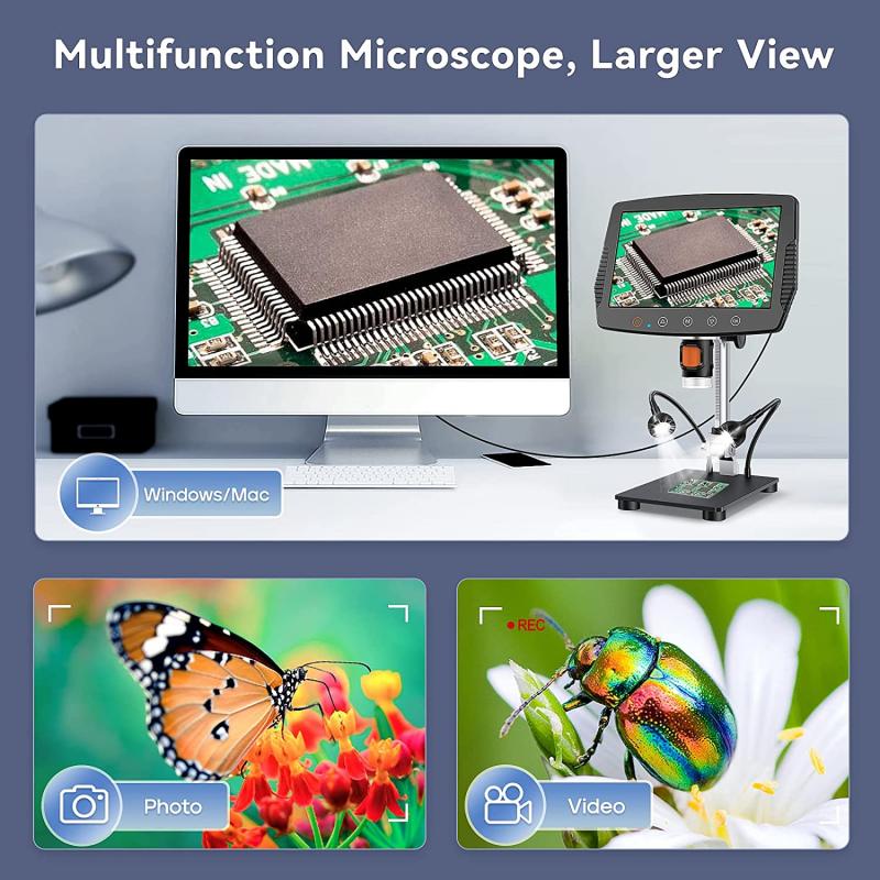
Types of Microscopes are used to magnify and observe objects that are too small to be seen with the naked eye. There are several types of microscopes available, each with its own unique features and applications. When looking through a microscope, there are several things to consider, including the type of microscope being used, the magnification level, and the quality of the optics.
One of the most common types of microscopes is the compound microscope, which is used to observe small, thin specimens such as cells, bacteria, and tissues. This type of microscope uses a series of lenses to magnify the specimen, and can be used to observe both living and non-living specimens.
Another type of microscope is the electron microscope, which uses a beam of electrons to magnify the specimen. This type of microscope is capable of much higher magnification levels than a compound microscope, and is often used to observe very small structures such as viruses and molecules.
Other types of microscopes include the stereo microscope, which is used to observe larger specimens such as insects and rocks, and the confocal microscope, which uses lasers to create a 3D image of the specimen.
When looking through a microscope, it is important to pay attention to the quality of the optics, as well as the lighting and focus. Proper lighting and focus can make a big difference in the clarity and detail of the image, and can help to identify important features of the specimen.
In recent years, advances in technology have led to the development of new types of microscopes, such as the super-resolution microscope, which is capable of observing structures at the molecular level. These new technologies are helping to push the boundaries of what we can observe and understand about the microscopic world.
4、 Illumination Methods

Illumination methods are an essential aspect of microscopy, as they determine the quality and clarity of the image produced. When looking at samples through a microscope, it is crucial to consider the illumination method used to ensure that the image produced is accurate and reliable.
One of the most common illumination methods used in microscopy is brightfield illumination. This method involves illuminating the sample with a bright light source from below, which allows for the observation of the sample's overall structure and morphology. However, this method may not be suitable for all samples, as it can result in low contrast and difficulty in distinguishing between different structures.
Another illumination method is darkfield illumination, which involves illuminating the sample with a hollow cone of light from the side. This method is particularly useful for observing samples that are transparent or have low contrast, as it enhances the visibility of the sample's edges and boundaries.
Fluorescence microscopy is another illumination method that involves the use of fluorescent dyes or proteins to label specific structures within the sample. This method allows for the observation of specific structures within the sample, such as proteins or DNA, and is widely used in biological research.
In recent years, super-resolution microscopy has emerged as a powerful tool for observing samples at the nanoscale level. This method involves the use of specialized illumination techniques, such as stimulated emission depletion (STED) microscopy or structured illumination microscopy (SIM), to achieve resolutions beyond the diffraction limit of light.
In conclusion, when looking at samples through a microscope, it is essential to consider the illumination method used to ensure that the image produced is accurate and reliable. Different illumination methods have their advantages and disadvantages, and the choice of method will depend on the sample being observed and the level of detail required. With the emergence of super-resolution microscopy, researchers can now observe samples at the nanoscale level, opening up new avenues for scientific discovery.


