When To Use Endoscope And Ultrasound ?
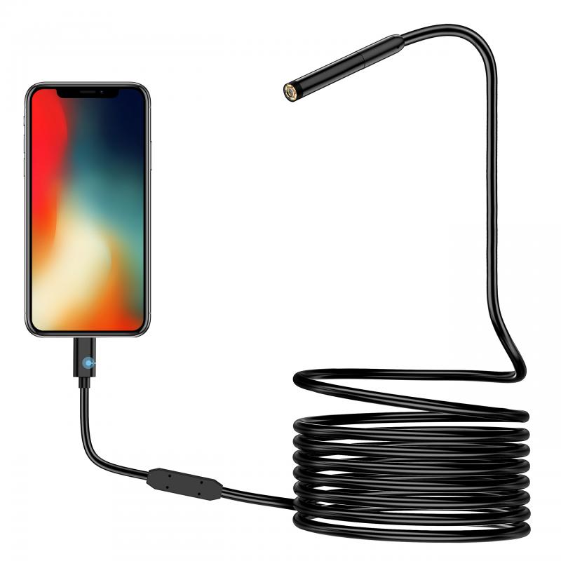
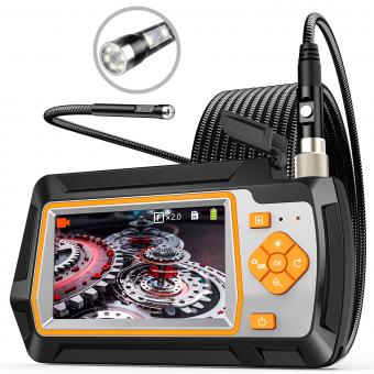
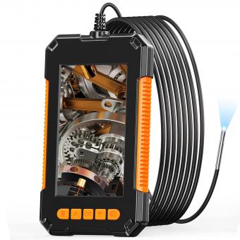
An endoscope is typically used for visualizing and examining the internal structures of the body, such as the gastrointestinal tract, respiratory system, or urinary system. It is a flexible or rigid tube with a light and camera attached to it, allowing doctors to see real-time images of the organs or tissues being examined. Endoscopy is commonly used for diagnosing and treating various conditions, including gastrointestinal disorders, respiratory diseases, and urological problems.
Ultrasound, on the other hand, uses high-frequency sound waves to create images of the body's internal structures. It is a non-invasive imaging technique that can be used to visualize organs, blood vessels, and soft tissues. Ultrasound is commonly used for monitoring fetal development during pregnancy, diagnosing and evaluating conditions affecting the abdomen, pelvis, or heart, and guiding certain medical procedures, such as biopsies or injections.
In summary, endoscopy is primarily used for direct visualization and examination of internal structures, while ultrasound is a non-invasive imaging technique used to create images of various body parts and guide medical procedures. The choice between the two depends on the specific medical condition or purpose of the examination.
1、 Endoscope: Applications in medical diagnosis and minimally invasive surgery.
Endoscopes and ultrasounds are both valuable tools in medical diagnosis and minimally invasive surgery. However, they are used in different scenarios and have distinct advantages.
Endoscopes are commonly used in the field of gastroenterology to visualize and diagnose conditions affecting the digestive system. They are inserted through natural body openings or small incisions and provide real-time images of the internal organs. Endoscopes can be used to detect and diagnose conditions such as ulcers, tumors, and gastrointestinal bleeding. They also allow for the collection of tissue samples for further analysis. In recent years, there have been advancements in endoscopic technology, such as high-definition imaging and the development of robotic-assisted endoscopy, which enhance the accuracy and precision of diagnosis and treatment.
On the other hand, ultrasounds use sound waves to create images of the body's internal structures. They are commonly used in obstetrics and gynecology to monitor fetal development and detect abnormalities. Ultrasounds are also used in cardiology to assess the heart's structure and function. Additionally, they are used in various other medical specialties, including urology, orthopedics, and radiology. Ultrasounds are non-invasive and do not involve radiation, making them safe for both patients and healthcare professionals.
The choice between endoscopes and ultrasounds depends on the specific medical condition and the area of the body being examined. Endoscopes are ideal for visualizing and diagnosing conditions within the digestive system, while ultrasounds are more versatile and can be used to examine various organs and structures. In some cases, both endoscopes and ultrasounds may be used together to provide a comprehensive evaluation of a patient's condition.
In conclusion, endoscopes and ultrasounds are valuable tools in medical diagnosis and minimally invasive surgery. The decision to use one over the other depends on the specific medical condition and the area of the body being examined. Both technologies continue to evolve, with advancements in imaging quality and robotic-assisted procedures, further enhancing their diagnostic and therapeutic capabilities.
2、 Endoscope: Advancements in imaging technology and therapeutic capabilities.
Endoscopes and ultrasound are both valuable tools in the field of medical imaging, but they serve different purposes and are used in different situations.
Endoscopes are used to visualize the internal structures of the body, typically through natural openings or small incisions. They are equipped with a light source and a camera, allowing doctors to examine areas such as the gastrointestinal tract, respiratory system, and urinary tract. Endoscopes have advanced significantly in recent years, with improvements in imaging technology and therapeutic capabilities. For example, high-definition cameras provide clearer and more detailed images, allowing for better diagnosis and treatment planning. Additionally, endoscopes can now be equipped with tools for minimally invasive procedures, such as removing polyps or performing biopsies.
Ultrasound, on the other hand, uses sound waves to create images of the body's internal structures. It is a non-invasive imaging technique that is commonly used to examine organs such as the heart, liver, and kidneys. Ultrasound is particularly useful for evaluating blood flow, as it can provide real-time images of the movement of blood through the vessels. It is also commonly used during pregnancy to monitor the development of the fetus.
The choice between endoscope and ultrasound depends on the specific medical condition and the area of the body being examined. Endoscopes are typically used when direct visualization of the internal structures is necessary, such as in the case of gastrointestinal disorders or respiratory conditions. Ultrasound, on the other hand, is often used for more general imaging purposes, such as evaluating organ function or monitoring fetal development.
In conclusion, endoscopes and ultrasound are both important tools in medical imaging, but they are used in different situations. Endoscopes are used for direct visualization of internal structures, while ultrasound is used for more general imaging purposes. The advancements in imaging technology and therapeutic capabilities of endoscopes have greatly improved their diagnostic and treatment capabilities.
3、 Ultrasound: Diagnostic imaging technique using high-frequency sound waves.
Endoscopy and ultrasound are both valuable diagnostic imaging techniques used in medical practice. While they serve different purposes, they can complement each other in certain situations.
Ultrasound is a non-invasive imaging technique that uses high-frequency sound waves to produce real-time images of the internal organs and structures of the body. It is commonly used to evaluate the abdomen, pelvis, heart, blood vessels, and musculoskeletal system. Ultrasound is particularly useful for visualizing soft tissues and fluid-filled structures, such as cysts or tumors. It is also safe and does not involve exposure to ionizing radiation, making it suitable for pregnant women and children. Additionally, recent advancements in ultrasound technology, such as 3D and 4D imaging, have further improved its diagnostic capabilities.
Endoscopy, on the other hand, involves the insertion of a flexible tube with a light and camera at its tip into the body to visualize the internal organs and structures directly. It is commonly used to examine the gastrointestinal tract, respiratory system, and urinary tract. Endoscopy allows for direct visualization of the mucosal lining, detection of abnormalities, and collection of tissue samples for biopsy. It is particularly useful for diagnosing conditions such as ulcers, polyps, tumors, and inflammation. However, endoscopy is an invasive procedure that may require sedation or anesthesia, and it carries a small risk of complications.
The choice between endoscopy and ultrasound depends on the specific clinical scenario. Ultrasound is often the initial imaging modality of choice due to its non-invasiveness and wide availability. It is used to screen for and diagnose a variety of conditions, such as gallstones, kidney stones, and abdominal aortic aneurysms. If an abnormality is detected on ultrasound, endoscopy may be recommended to further evaluate and obtain a more detailed assessment. For example, if an ultrasound detects a suspicious mass in the gastrointestinal tract, an endoscopy can be performed to directly visualize the lesion and obtain a biopsy for definitive diagnosis.
In summary, ultrasound and endoscopy are both valuable diagnostic tools in medical practice. Ultrasound is non-invasive, safe, and widely used for initial screening and evaluation of various conditions. Endoscopy, on the other hand, allows for direct visualization and biopsy of internal structures, making it useful for further evaluation and diagnosis of abnormalities detected on ultrasound. The choice between the two depends on the specific clinical scenario and the information needed for accurate diagnosis and treatment.

4、 Ultrasound: Applications in obstetrics, cardiology, and musculoskeletal imaging.
Endoscopy and ultrasound are both valuable diagnostic tools used in various medical fields. While endoscopy involves the use of a flexible or rigid tube with a light and camera to visualize internal organs and structures, ultrasound utilizes high-frequency sound waves to create images of the body's internal structures.
Ultrasound is commonly used in obstetrics to monitor the development and health of the fetus during pregnancy. It allows healthcare professionals to assess fetal growth, detect abnormalities, and evaluate the placenta and amniotic fluid. In cardiology, ultrasound, also known as echocardiography, is used to assess the structure and function of the heart, including the valves, chambers, and blood flow. It helps in diagnosing conditions such as heart defects, heart failure, and heart valve abnormalities.
In musculoskeletal imaging, ultrasound is used to evaluate soft tissues, muscles, tendons, ligaments, and joints. It aids in diagnosing conditions like tendonitis, bursitis, sprains, and tears. Ultrasound-guided procedures, such as joint injections or biopsies, are also performed to enhance accuracy and safety.
The latest point of view regarding ultrasound is the advancement in technology, such as the development of 3D and 4D ultrasound imaging. These techniques provide more detailed and realistic images, allowing for better visualization and diagnosis. Additionally, portable ultrasound devices have become more accessible, enabling point-of-care imaging in various clinical settings.
On the other hand, endoscopy is used when direct visualization of internal organs or structures is required. It is commonly used in gastroenterology to examine the gastrointestinal tract, including the esophagus, stomach, and colon. Endoscopy allows for the detection and diagnosis of conditions such as ulcers, polyps, and tumors. It is also used in urology, gynecology, and pulmonology to visualize and diagnose conditions specific to those areas.
In summary, ultrasound is primarily used in obstetrics, cardiology, and musculoskeletal imaging, providing non-invasive imaging of internal structures. Endoscopy, on the other hand, is used when direct visualization is necessary, primarily in gastroenterology, urology, gynecology, and pulmonology. Both techniques have their unique applications and play crucial roles in diagnosing and managing various medical conditions.















![J12 Mini-projector Outdoor-filmprojector met 100 inch-projectorscherm, 1080P, compatibel met tv-stick, videogames, HDMI, USB, TF, VGA, AUX, AV [Amerikaanse regelgeving] J12 Mini-projector Outdoor-filmprojector met 100 inch-projectorscherm, 1080P, compatibel met tv-stick, videogames, HDMI, USB, TF, VGA, AUX, AV [Amerikaanse regelgeving]](https://img.kentfaith.de/cache/catalog/products/de/GW01.0172/GW01.0172-1-200x200.jpg)


