When Using A Microscope ?
When using a microscope, it is important to handle the instrument with care and follow proper procedures. This includes ensuring that the microscope is clean and free from dust or debris, as this can affect the quality of the images. Additionally, it is crucial to adjust the focus and lighting settings to obtain a clear and magnified view of the specimen. Proper positioning of the slide and using the appropriate objective lens are also essential for obtaining accurate results. Finally, it is important to record observations and findings accurately, as well as clean and store the microscope properly after use to maintain its longevity.
1、 Magnification
When using a microscope, magnification is a crucial aspect that allows scientists and researchers to observe and study objects at a much larger scale than what is visible to the naked eye. Magnification refers to the increase in apparent size of an object when viewed through a microscope.
Microscopes have evolved significantly over the years, with advancements in technology leading to higher magnification capabilities. Traditional light microscopes use lenses to magnify the image, while electron microscopes use beams of electrons to achieve even higher magnification.
Magnification plays a vital role in various scientific fields. In biology, for example, it enables the study of cellular structures, such as organelles within cells, and the observation of microscopic organisms. In materials science, magnification allows for the examination of the atomic and molecular structure of materials, aiding in the development of new materials with specific properties.
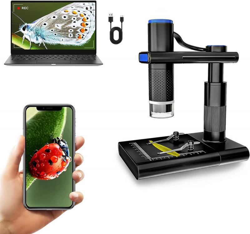
The latest point of view regarding magnification is the increasing use of digital microscopes. These microscopes utilize digital imaging technology to capture images and videos of the specimen, which can then be viewed on a computer screen or other digital devices. Digital microscopes often have built-in software that allows for image analysis and measurement, providing researchers with more accurate and efficient data.
Furthermore, advancements in artificial intelligence and machine learning have enabled automated image analysis, making it easier to process and interpret large amounts of data obtained through magnification. This has opened up new possibilities for research and discovery in various fields.
In conclusion, magnification is a fundamental aspect of microscopy that allows scientists to explore the microscopic world. With the latest advancements in technology, such as digital microscopes and automated image analysis, magnification continues to play a crucial role in scientific research and discovery.

2、 Resolution
When using a microscope, resolution refers to the ability of the microscope to distinguish two closely spaced objects as separate entities. It is a measure of the level of detail that can be observed and is an important factor in microscopy.
Resolution is determined by the numerical aperture of the microscope's objective lens and the wavelength of light used. The numerical aperture is a measure of the lens's ability to gather light and is influenced by the lens's design and the refractive index of the medium between the lens and the specimen. The shorter the wavelength of light used, the higher the resolution that can be achieved.
In recent years, there have been advancements in microscopy techniques that have pushed the limits of resolution beyond what was previously thought possible. One such technique is super-resolution microscopy, which allows for the visualization of structures that are smaller than the diffraction limit of light. This is achieved by using fluorescent molecules that can be switched on and off, allowing for the precise localization of individual molecules. Super-resolution microscopy has revolutionized the field of cell biology, enabling researchers to study cellular structures and processes at an unprecedented level of detail.
Additionally, advancements in electron microscopy have also contributed to improved resolution. Electron microscopes use a beam of electrons instead of light, which has a much shorter wavelength, allowing for higher resolution imaging. Techniques such as cryo-electron microscopy have further enhanced the resolution of electron microscopy, enabling the visualization of biological structures at near-atomic resolution.
In conclusion, when using a microscope, resolution is a critical factor in determining the level of detail that can be observed. Recent advancements in microscopy techniques, such as super-resolution microscopy and cryo-electron microscopy, have pushed the limits of resolution and revolutionized our understanding of the microscopic world.
3、 Illumination
When using a microscope, illumination is a crucial aspect that greatly impacts the quality and clarity of the observed specimen. Illumination refers to the light source that is used to illuminate the specimen being viewed under the microscope. It plays a vital role in enhancing contrast, resolution, and overall visibility of the specimen.
Traditionally, microscopes used to rely on external light sources such as natural light or lamps. However, with advancements in technology, modern microscopes now incorporate built-in LED or halogen light sources. These light sources provide a consistent and adjustable illumination, allowing for better control and optimization of the viewing conditions.
The latest point of view regarding illumination in microscopy emphasizes the importance of using appropriate lighting techniques to enhance the visualization of specific structures within the specimen. For example, techniques like darkfield and phase contrast microscopy utilize specialized lighting methods to improve the visibility of transparent or low-contrast specimens. These techniques enable researchers to observe fine details that may otherwise be difficult to detect.
Furthermore, advancements in fluorescence microscopy have revolutionized the field of biological imaging. Fluorescent dyes and markers, combined with specific illumination techniques, allow for the visualization of specific molecules or structures within living cells. This has opened up new avenues for studying cellular processes and disease mechanisms.
In conclusion, illumination is a critical factor when using a microscope as it directly impacts the quality of observation. The latest advancements in lighting techniques have significantly improved the visualization of specimens, enabling researchers to explore the microscopic world with greater precision and detail.

4、 Focus
When using a microscope, focus is a crucial aspect that determines the clarity and sharpness of the observed specimen. Achieving proper focus allows scientists and researchers to examine the intricate details of microscopic structures and gain a deeper understanding of the world at a cellular level.
To focus a microscope, one must adjust the position of the objective lens or lenses to bring the specimen into sharp focus. This is typically done by turning the coarse focus knob to initially bring the specimen into view, and then fine-tuning the focus using the fine focus knob for precise clarity. The process requires patience and careful adjustments to ensure the best possible image quality.
In recent years, advancements in microscope technology have led to the development of new focusing techniques. One such technique is called autofocus, which uses sensors and algorithms to automatically adjust the focus of the microscope. This is particularly useful when observing live specimens or when capturing time-lapse images, as it eliminates the need for constant manual adjustments.
Additionally, the introduction of digital microscopes has revolutionized the way focus is achieved. These microscopes use digital imaging sensors and software to capture images, allowing for post-capture focus adjustments. This means that even if the initial focus was not perfect, researchers can still obtain clear images by adjusting the focus digitally after the image is captured.
In conclusion, focus is a fundamental aspect of using a microscope. Whether through manual adjustments or the latest autofocus and digital imaging technologies, achieving proper focus is essential for obtaining clear and detailed microscopic images. These advancements continue to enhance the capabilities of microscopes and contribute to the progress of scientific research and discovery.


