Where Are Slides Placed On A Microscope ?
Slides are placed on a microscope stage.
1、 "Microscope Slide Placement: Positioning on the Stage"
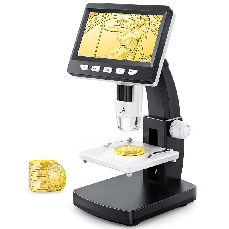
Microscope slides are placed on the stage of a microscope. The stage is a flat platform that holds the specimen being observed. It is located beneath the objective lenses and above the light source. The stage typically has a mechanical stage control that allows for precise movement of the slide in both the x and y directions.
When placing a microscope slide on the stage, it is important to ensure that the slide is centered and properly aligned. This is crucial for obtaining clear and accurate images. The slide should be positioned in such a way that the area of interest is directly under the objective lens. This can be achieved by using the mechanical stage control to move the slide until the desired area is in the center of the field of view.
In recent years, there have been advancements in microscope technology that have introduced automated slide placement systems. These systems use computer algorithms to analyze the slide and automatically position it on the stage. This eliminates the need for manual adjustment and ensures consistent and accurate positioning of the slide.
Additionally, some microscopes now come with digital imaging capabilities, allowing for the capture and analysis of images directly on a computer. In these cases, the slide may be placed on a separate digital imaging stage that is connected to the microscope. This allows for easy transfer of images and facilitates further analysis and sharing of data.
In conclusion, microscope slides are placed on the stage of a microscope. Proper positioning and alignment of the slide are essential for obtaining clear and accurate images. Advancements in technology have introduced automated slide placement systems and digital imaging capabilities, further enhancing the efficiency and accuracy of microscope slide placement.

2、 "Proper Placement of Slides on a Microscope Stage"
The proper placement of slides on a microscope stage is crucial for obtaining clear and accurate images. The stage is the platform on which the slide is placed, and it is designed to hold the slide securely in place while allowing it to be moved in different directions for observation.
Typically, the slide is placed on the stage with the specimen facing upwards. The slide should be centered on the stage, ensuring that the specimen is directly under the objective lens. This allows the light to pass through the specimen and be magnified by the lens, resulting in a clear image.
In recent years, advancements in microscope technology have led to the development of motorized stages. These stages can automatically position the slide, making it easier for researchers to locate and focus on specific areas of interest. Additionally, some microscopes now have digital imaging capabilities, allowing users to capture and analyze images directly on a computer screen.
It is important to note that different types of microscopes may have slightly different stage designs. For example, inverted microscopes have a stage that is located above the objective lens, and the slide is placed upside down on the stage. This configuration is commonly used in cell culture and live cell imaging applications.
In conclusion, the proper placement of slides on a microscope stage is essential for obtaining accurate and high-quality images. Researchers should ensure that the slide is centered on the stage and that the specimen is directly under the objective lens. Advancements in microscope technology, such as motorized stages and digital imaging capabilities, have made slide placement and observation more efficient and convenient.

3、 "Understanding the Location of Slides on a Microscope"
Slides are an essential component of a microscope as they hold the specimen that is being observed. The location of slides on a microscope is crucial for proper viewing and analysis. Typically, slides are placed on the stage of the microscope, which is a flat platform that holds the specimen in position for examination.
The stage is located beneath the objective lenses and above the light source. It is usually equipped with clips or clamps to secure the slide in place. The slide is positioned on the stage with the specimen facing upward, allowing light to pass through it for magnification and observation.
The stage is designed to be adjustable, allowing the user to move the slide horizontally and vertically to position the area of interest under the objective lenses. This adjustability is important for focusing and obtaining a clear image of the specimen.
In recent years, advancements in microscope technology have led to the development of motorized stages. These stages can be controlled electronically, allowing for precise movement and automation of slide positioning. This feature has greatly improved the efficiency and accuracy of microscopic analysis, particularly in fields such as pathology and research.
In conclusion, slides are placed on the stage of a microscope, which is located beneath the objective lenses. The stage is adjustable to position the specimen for optimal viewing. With the advent of motorized stages, slide positioning has become more precise and automated, enhancing the capabilities of microscopic analysis.
4、 "Slide Positioning: A Crucial Step in Microscopy"
Slides are an essential component of a microscope, as they hold the specimen that is being observed. The positioning of slides on a microscope is a crucial step in microscopy, as it directly affects the quality of the observation and the accuracy of the results obtained.
Traditionally, slides are placed on the stage of the microscope. The stage is a flat platform that holds the slide in place and allows for precise movement and positioning. The slide is typically secured on the stage using clips or clamps to prevent any movement during observation. This ensures that the specimen remains in focus and allows for easy manipulation of the slide when changing the field of view.
However, with advancements in microscopy technology, there have been some changes in slide positioning. For example, some modern microscopes now have motorized stages that can automatically move the slide in different directions, allowing for more efficient scanning of large specimens or multiple regions of interest. Additionally, some microscopes now have slide holders or trays that can hold multiple slides simultaneously, enabling quick and easy switching between different specimens.
Furthermore, the introduction of digital microscopy has revolutionized slide positioning. Digital microscopes allow for the capture of high-resolution images or videos of the specimen, which can be viewed and analyzed on a computer screen. In this case, the slide may be placed on a specialized digital microscope scanner or a slide scanner, which automatically moves the slide and captures multiple images at different focal planes. This allows for the creation of a detailed, three-dimensional representation of the specimen.
In conclusion, the positioning of slides on a microscope is a crucial step in microscopy. While traditionally slides are placed on the stage, advancements in technology have introduced motorized stages, slide holders, and digital microscopy, which have expanded the possibilities for slide positioning and improved the efficiency and accuracy of observations.
