Which Microscope Uses Visible Light ?
A light microscope, also known as an optical microscope, uses visible light to illuminate the specimen being observed. This type of microscope is the most commonly used in biology and medicine, as it allows for the observation of living cells and tissues. Light microscopes can magnify specimens up to 1000 times their original size, and can also be used to observe the internal structures of cells and tissues. The basic components of a light microscope include a light source, lenses, and a stage for holding the specimen. The lenses in a light microscope can be adjusted to change the magnification and focus of the image. In addition to brightfield microscopy, which is the most common type of light microscopy, there are also other types of light microscopy, such as fluorescence microscopy and confocal microscopy, which use specialized techniques to enhance the contrast and resolution of the image.
1、 Compound light microscope
The compound light microscope is the type of microscope that uses visible light to produce an image of a specimen. This type of microscope is widely used in various fields such as biology, medicine, and materials science. It is a versatile and powerful tool that allows scientists to observe and study the structure and behavior of microscopic organisms and materials.
The compound light microscope works by passing visible light through a specimen and then through a series of lenses that magnify the image. The lenses in a compound light microscope are arranged in a series of two or more, which allows for higher magnification and resolution than a single lens microscope. The image produced by a compound light microscope is two-dimensional and can be viewed directly through the eyepiece or captured using a camera.
In recent years, there has been a growing interest in developing new types of microscopes that use different types of light, such as infrared or ultraviolet, to produce images of specimens. These new types of microscopes offer unique advantages over traditional compound light microscopes, such as the ability to see through opaque materials or to study the chemical composition of a specimen. However, the compound light microscope remains the most widely used type of microscope in many fields due to its versatility, ease of use, and relatively low cost.

2、 Stereo microscope
Which microscope uses visible light? The stereo microscope is one of the most commonly used microscopes that utilizes visible light. It is also known as a dissecting microscope and is used to view larger specimens that cannot be viewed under a compound microscope. The stereo microscope uses two separate optical paths with two eyepieces, which provides a three-dimensional view of the specimen. It has a lower magnification range compared to a compound microscope, typically ranging from 10x to 40x magnification.
The stereo microscope is widely used in various fields such as biology, geology, and electronics. In biology, it is used to study the external features of organisms, such as insects, plants, and small animals. In geology, it is used to examine rocks, minerals, and fossils. In electronics, it is used to inspect circuit boards and other small components.
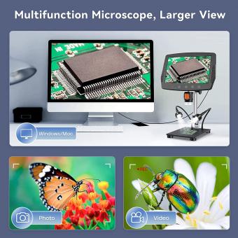
Advancements in technology have led to the development of digital stereo microscopes, which allow for the capture of high-resolution images and videos of the specimen. These microscopes also have the ability to connect to a computer or other devices for further analysis and sharing of data.
In conclusion, the stereo microscope is a valuable tool in various fields that uses visible light to provide a three-dimensional view of the specimen. With the latest advancements in technology, it has become even more versatile and useful in research and analysis.
3、 Polarizing microscope
Which microscope uses visible light? The answer is a polarizing microscope. This type of microscope uses polarized light to examine samples. The polarized light is passed through a polarizer, which only allows light waves to pass through in a specific orientation. The light then passes through the sample, which may cause the light waves to change orientation. The light then passes through an analyzer, which only allows light waves to pass through in a specific orientation that is perpendicular to the polarizer. The resulting image shows the areas of the sample that caused the light waves to change orientation.
Polarizing microscopes are commonly used in geology, materials science, and biology. In geology, they are used to examine the optical properties of minerals. In materials science, they are used to examine the structure of materials, such as polymers and crystals. In biology, they are used to examine the structure of cells and tissues.
Recent advances in polarizing microscopy have led to the development of new techniques, such as polarized light microscopy and polarized fluorescence microscopy. These techniques allow for the visualization of specific molecules and structures within cells and tissues. They have also been used to study the properties of materials, such as liquid crystals and nanomaterials.
In conclusion, polarizing microscopes use visible light to examine samples and are commonly used in geology, materials science, and biology. Recent advances in polarizing microscopy have led to the development of new techniques that allow for the visualization of specific molecules and structures within cells and tissues.
4、 Fluorescence microscope
Which microscope uses visible light? The answer is the fluorescence microscope. This type of microscope uses visible light to excite fluorescent molecules in a sample, which then emit light of a different color that can be detected and visualized. Fluorescence microscopy has become an essential tool in many fields of science, including biology, medicine, and materials science.
One of the latest developments in fluorescence microscopy is super-resolution microscopy, which allows for imaging at a resolution beyond the diffraction limit of light. This has enabled researchers to visualize structures and processes at the nanoscale level, providing new insights into cellular and molecular biology.
Another recent advancement in fluorescence microscopy is the use of genetically encoded fluorescent proteins, which can be expressed in living cells and organisms to label specific structures or molecules. This has revolutionized the field of cell biology, allowing for dynamic imaging of cellular processes in real-time.
Overall, fluorescence microscopy has become an indispensable tool for researchers in many fields, and its continued development and refinement will undoubtedly lead to new discoveries and insights in the years to come.


