Which Type Of Microscope ?
There are several types of microscopes, including optical microscopes, electron microscopes, and scanning probe microscopes. The type of microscope used depends on the specific application and the level of magnification required. Optical microscopes use visible light to magnify specimens and are commonly used in biology and medicine. Electron microscopes use beams of electrons to create high-resolution images and are often used in materials science and nanotechnology. Scanning probe microscopes use a physical probe to scan the surface of a sample and are used in fields such as chemistry and physics.
1、 Compound Microscope
Which type of microscope is best for observing small, transparent specimens such as cells and bacteria? The answer is a Compound Microscope. This type of microscope uses two or more lenses to magnify the specimen, allowing for a detailed view of its internal structures. Compound microscopes are commonly used in biology, medicine, and research laboratories.
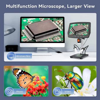
In recent years, advancements in technology have led to the development of digital compound microscopes. These microscopes use a camera to capture images of the specimen, which can be viewed on a computer screen or saved for later analysis. Digital microscopes have become increasingly popular in education and research, as they allow for easy sharing and collaboration among scientists and students.
Another recent development in compound microscopy is the use of fluorescence. Fluorescence microscopy uses fluorescent dyes to label specific structures within the specimen, allowing for a more detailed view of its internal workings. This technique has revolutionized the study of cellular processes and has led to many important discoveries in the field of biology.
Overall, the compound microscope remains an essential tool in the study of small, transparent specimens. With the latest advancements in technology, this type of microscope continues to evolve and improve, allowing scientists to gain a deeper understanding of the microscopic world around us.

2、 Electron Microscope
Which type of microscope is best for observing extremely small structures such as viruses and bacteria? The answer is an Electron Microscope. Unlike light microscopes, which use visible light to magnify objects, electron microscopes use a beam of electrons to create an image. This allows for much higher magnification and resolution, making it possible to see structures as small as a few nanometers.
There are two main types of electron microscopes: Transmission Electron Microscopes (TEM) and Scanning Electron Microscopes (SEM). TEMs use a thin sample that is placed on a grid and then bombarded with electrons. The electrons pass through the sample and create an image on a screen. SEMs, on the other hand, use a beam of electrons to scan the surface of a sample and create a 3D image.
The latest advancements in electron microscopy include the development of cryo-electron microscopy (cryo-EM), which allows for the imaging of biological molecules in their natural state. This technique involves freezing the sample in liquid nitrogen and then imaging it at very low temperatures. Cryo-EM has revolutionized the field of structural biology, allowing scientists to study the structure of proteins and other biomolecules at atomic resolution.
In conclusion, electron microscopes are the best type of microscope for observing extremely small structures such as viruses and bacteria. The latest advancements in electron microscopy, such as cryo-EM, have opened up new avenues for research in the field of structural biology.

3、 Scanning Probe Microscope
Which type of microscope is a Scanning Probe Microscope (SPM). SPM is a type of microscope that uses a physical probe to scan the surface of a sample to create an image with high resolution. The probe is usually a sharp tip that is scanned across the surface of the sample, and the interaction between the tip and the sample is measured to create an image.
SPM has become an important tool in nanotechnology research because it allows scientists to study the properties of materials at the nanoscale. SPM can be used to measure the topography, conductivity, and magnetic properties of materials with high resolution. This makes it an important tool for studying the properties of materials used in electronics, medicine, and other fields.
One of the latest developments in SPM is the use of machine learning algorithms to analyze the data collected by the microscope. Machine learning algorithms can be used to identify patterns in the data that are difficult for humans to detect. This can help scientists to better understand the properties of materials at the nanoscale and to develop new materials with specific properties.
Overall, SPM is an important tool for studying the properties of materials at the nanoscale. The latest developments in machine learning algorithms are making it even more powerful and useful for scientific research.

4、 Confocal Microscope
Which type of microscope is a Confocal Microscope?
A Confocal Microscope is a type of microscope that uses a laser beam to scan a sample and create a three-dimensional image. It is a powerful tool for studying biological samples, as it allows researchers to visualize the internal structures of cells and tissues with high resolution and clarity.
One of the key advantages of a Confocal Microscope is its ability to eliminate out-of-focus light, which results in sharper images and better contrast. This is achieved through a pinhole aperture that only allows light from a specific focal plane to pass through, while blocking light from other planes.
In recent years, Confocal Microscopy has become an increasingly popular technique in the field of neuroscience, as it allows researchers to study the complex structures of the brain in great detail. It has also been used in the study of cancer, where it has helped to identify new targets for drug development.
Overall, the Confocal Microscope is a powerful tool for studying biological samples, and its use is likely to continue to grow in the coming years as researchers seek to better understand the complex structures and processes of living organisms.


