Who Created The Light Microscope ?
The light microscope was not created by a single individual, but rather developed over time by multiple scientists. The earliest known microscope was created by Dutch spectacle maker Zacharias Janssen in the late 16th century. However, it was not until the 17th century that the microscope was refined and improved by scientists such as Robert Hooke and Antonie van Leeuwenhoek. Hooke is credited with coining the term "cell" after observing the structure of cork under a microscope, while van Leeuwenhoek is known for his extensive observations of microorganisms. The development of the light microscope revolutionized the study of biology and medicine, allowing scientists to observe and study the microscopic world in greater detail.
1、 Invention of the Light Microscope
The light microscope was invented by Dutch scientist Antonie van Leeuwenhoek in the late 17th century. He is often referred to as the "father of microbiology" for his pioneering work in observing and documenting microscopic organisms, including bacteria and protozoa. Leeuwenhoek's microscope consisted of a single lens, which he ground himself, and was capable of magnifying objects up to 300 times their original size.
However, it is important to note that the invention of the light microscope was not a single event, but rather a gradual development over several centuries. The first known microscope was created in the early 1600s by Dutch spectacle makers Hans and Zacharias Janssen, who used multiple lenses to magnify objects. This was followed by the compound microscope, which used two or more lenses to produce a more detailed image.
In recent years, there has been some debate over the true inventor of the light microscope. Some historians have suggested that Italian scientist Giuseppe Campani may have invented a similar microscope before Leeuwenhoek, but there is limited evidence to support this claim.
Regardless of who can be credited with the invention of the light microscope, there is no denying its profound impact on science and medicine. Today, microscopes are used in a wide range of fields, from biology and chemistry to materials science and engineering. They have allowed us to explore and understand the microscopic world in ways that were once unimaginable.

2、 Early Development of the Light Microscope
The credit for creating the first light microscope is often given to Dutch scientist Antonie van Leeuwenhoek in the late 17th century. He was able to achieve magnifications of up to 270 times using a simple microscope with a single lens. However, it is important to note that the development of the light microscope was a gradual process that involved the contributions of many scientists over several centuries.
In the early 1600s, Galileo Galilei and Johannes Kepler developed the concept of using lenses to magnify objects. Later, in the mid-1600s, Robert Hooke and Marcello Malpighi used compound microscopes with multiple lenses to study biological specimens. However, it was Leeuwenhoek who made significant advancements in the field by perfecting the single-lens microscope and using it to observe microorganisms and other tiny structures.
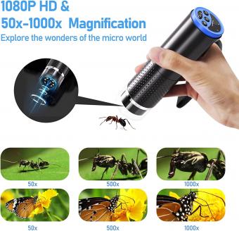
Today, the light microscope remains an essential tool in the field of biology, allowing scientists to study cells, tissues, and microorganisms in detail. However, modern microscopes have undergone significant technological advancements, including the use of digital imaging and advanced optics, which have greatly improved their capabilities.
In conclusion, while Antonie van Leeuwenhoek is often credited with creating the first light microscope, the development of this essential scientific tool was a gradual process that involved the contributions of many scientists over several centuries. Today, the light microscope continues to play a crucial role in scientific research and discovery.

3、 Improvements in Light Microscope Design
The light microscope was invented in the 17th century, and its creation is attributed to Dutch scientist Antonie van Leeuwenhoek. However, improvements in light microscope design have been made over the years by various scientists and inventors.
One of the most significant improvements in light microscope design was made by German physicist Ernst Abbe in the late 19th century. Abbe developed a mathematical formula that allowed for the correction of chromatic and spherical aberrations in microscope lenses, resulting in clearer and sharper images.
In the 20th century, the development of electron microscopy allowed for even greater magnification and resolution of biological specimens. However, light microscopy remains an important tool in biological research, particularly for studying living cells and tissues.
Recent advancements in light microscopy include the development of super-resolution microscopy techniques, which allow for imaging at the nanoscale level. These techniques have revolutionized our understanding of cellular structures and processes, and have led to new discoveries in fields such as neuroscience and immunology.
Overall, the light microscope has undergone significant improvements in design since its invention, and continues to be a valuable tool in biological research.

4、 Applications of the Light Microscope in Science
Who created the light microscope?
The credit for creating the first light microscope goes to Dutch scientist Antonie van Leeuwenhoek in the late 17th century. He used a simple microscope with a single lens to observe and study microorganisms, which he called "animalcules."
Applications of the Light Microscope in Science:
The light microscope has been a crucial tool in scientific research for centuries. It has allowed scientists to observe and study the structure and function of cells, tissues, and organisms at the microscopic level. Some of the key applications of the light microscope in science include:
1. Cell Biology: The light microscope has been instrumental in advancing our understanding of cell biology. It has allowed scientists to observe and study the structure and function of cells, including their organelles, cytoskeleton, and membrane systems.
2. Microbiology: The light microscope has been used extensively in microbiology to study microorganisms such as bacteria, viruses, and fungi. It has allowed scientists to observe and study the morphology, structure, and behavior of these microorganisms.
3. Histology: The light microscope has been used in histology to study the structure and function of tissues. It has allowed scientists to observe and study the different types of cells that make up tissues, as well as their organization and function.
4. Medical Diagnosis: The light microscope has been used in medical diagnosis to study blood cells, urine samples, and other bodily fluids. It has allowed doctors to diagnose diseases and conditions based on the presence or absence of certain cells or microorganisms.
5. Material Science: The light microscope has been used in material science to study the structure and properties of materials at the microscopic level. It has allowed scientists to observe and study the microstructure of materials, including metals, ceramics, and polymers.
In recent years, advances in technology have led to the development of new types of microscopes, such as the electron microscope and the confocal microscope. However, the light microscope remains a crucial tool in scientific research, and its applications continue to expand as new techniques and technologies are developed.


