Why Vets Use Endoscope When Placing Peg Tubes ?
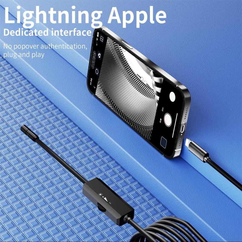
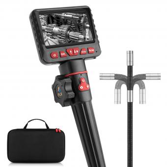
Veterinarians use endoscopes when placing percutaneous endoscopic gastrostomy (PEG) tubes in animals because it allows for a minimally invasive procedure. The endoscope is a flexible tube with a camera and light source that can be inserted through a small incision or natural body opening. By using an endoscope, vets can visualize the internal organs and guide the placement of the PEG tube without the need for extensive surgery. This reduces the risk of complications, minimizes pain and discomfort for the animal, and promotes faster recovery. Additionally, the endoscope allows vets to monitor the placement of the tube in real-time, ensuring accurate positioning and reducing the likelihood of complications such as perforation or dislodgement. Overall, the use of endoscopes in PEG tube placement is a safe and effective technique that benefits both the veterinarian and the animal.
1、 Visualization: Assessing the internal structures for accurate tube placement.
Vets use endoscopes when placing peg tubes for several reasons, with visualization being one of the primary factors. Endoscopy allows vets to assess the internal structures of the patient's gastrointestinal tract, ensuring accurate tube placement.
By inserting an endoscope into the patient's body, vets can visualize the esophagus, stomach, and duodenum, among other structures. This visualization helps them identify any abnormalities or potential complications that may affect the placement of the peg tube. For example, they can identify strictures, tumors, or other obstructions that could hinder the tube's proper positioning.
Accurate tube placement is crucial to ensure the effectiveness and safety of the peg tube. Improper placement can lead to complications such as aspiration pneumonia, leakage, or dislodgement. By using an endoscope, vets can visually confirm that the tube is correctly positioned within the stomach or small intestine, minimizing the risk of these complications.
Moreover, the use of endoscopy allows vets to make real-time adjustments during the procedure. They can manipulate the endoscope to obtain different angles and perspectives, ensuring optimal visualization and precise tube placement. This dynamic approach enhances the vet's ability to navigate through the gastrointestinal tract and overcome any challenges that may arise.
It is important to note that the latest point of view in veterinary medicine emphasizes the importance of minimally invasive techniques. Endoscopy offers a less invasive alternative to traditional surgical procedures, reducing patient discomfort, recovery time, and the risk of complications. As a result, the use of endoscopes for peg tube placement has become increasingly common in veterinary practice.
In conclusion, vets use endoscopes when placing peg tubes primarily for visualization purposes. Assessing the internal structures of the gastrointestinal tract allows vets to accurately position the tube, minimizing the risk of complications and improving patient outcomes. The latest trend in veterinary medicine supports the use of minimally invasive techniques, making endoscopy an increasingly popular choice for peg tube placement.
2、 Minimally invasive: Reducing the need for extensive surgical procedures.
Vets use endoscopes when placing peg tubes in animals for several reasons, with the primary one being the minimally invasive nature of the procedure. Endoscopy allows veterinarians to perform the placement of peg tubes without the need for extensive surgical procedures. This means that the animal experiences less trauma, pain, and a faster recovery time.
Endoscopy involves the use of a flexible tube with a camera and light source at the end, which is inserted into the animal's body through a small incision or natural opening. The camera provides real-time images of the internal organs, allowing the vet to guide the placement of the peg tube accurately. This minimally invasive approach reduces the risk of complications, such as infection and bleeding, and also decreases the length of hospital stays for the animals.
Furthermore, endoscopy offers veterinarians a better view of the internal structures, allowing for more precise placement of the peg tube. This is particularly important in cases where the animal has underlying health conditions or anatomical abnormalities that may complicate the procedure. By using endoscopy, vets can ensure that the peg tube is correctly positioned, minimizing the risk of complications and improving the overall success of the procedure.
From the latest point of view, advancements in endoscopic technology have further improved the effectiveness of peg tube placement. For example, the development of smaller and more flexible endoscopes allows for easier access to the animal's gastrointestinal tract, reducing the need for larger incisions. Additionally, the integration of high-definition imaging and advanced surgical tools into endoscopic systems enables vets to perform more complex procedures with greater precision.
In conclusion, vets use endoscopes when placing peg tubes in animals primarily because it is a minimally invasive procedure. This approach reduces the need for extensive surgical procedures, minimizes trauma and pain for the animals, and promotes faster recovery. With the continuous advancements in endoscopic technology, the effectiveness and precision of peg tube placement have significantly improved, benefiting both the animals and veterinary professionals.
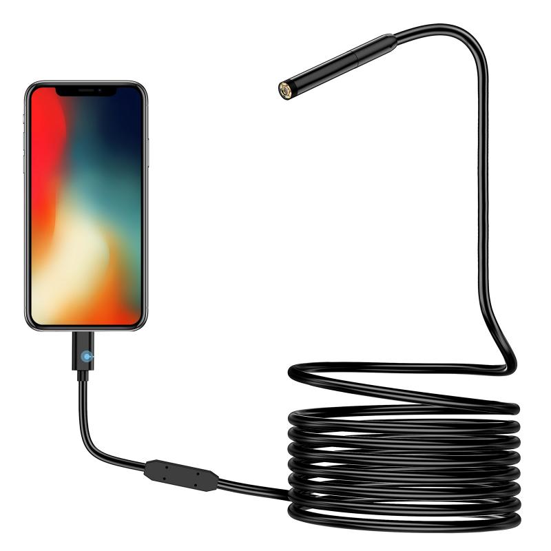
3、 Precision: Ensuring precise and targeted tube insertion.
Vets use endoscopes when placing peg tubes for several reasons, with precision being one of the key factors. Endoscopes are medical devices that consist of a long, flexible tube with a light and camera attached to it. This allows veterinarians to visualize the internal organs and structures of animals without the need for invasive surgery.
When placing peg tubes, precision is crucial to ensure accurate and targeted tube insertion. The endoscope provides a clear view of the gastrointestinal tract, allowing vets to identify the optimal location for tube placement. This is particularly important in cases where the animal has specific medical conditions or anatomical abnormalities that require careful consideration.
By using an endoscope, vets can navigate through the esophagus and stomach with precision, avoiding any potential complications or injuries. They can visualize the exact position of the tube and ensure it is correctly placed in the stomach or small intestine. This precision minimizes the risk of complications such as leakage, dislodgement, or damage to surrounding tissues.
Moreover, the use of endoscopes in peg tube placement has evolved with advancements in technology. The latest endoscopic equipment provides high-definition imaging, allowing vets to visualize even the smallest details with exceptional clarity. This enhanced visualization aids in accurate tube placement and reduces the chances of errors or complications.
In conclusion, vets use endoscopes when placing peg tubes primarily for precision. The ability to visualize the gastrointestinal tract and accurately position the tube is crucial for successful tube insertion. With the latest advancements in endoscopic technology, vets can ensure precise and targeted tube placement, improving the overall outcome for the animal.
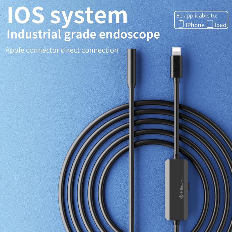
4、 Diagnostic capabilities: Identifying any underlying conditions or abnormalities.
Vets use endoscopes when placing peg tubes for several reasons, with one of the main reasons being the diagnostic capabilities that this procedure offers. Endoscopy allows veterinarians to visually examine the gastrointestinal tract and identify any underlying conditions or abnormalities that may be present.
By inserting an endoscope into the animal's body, the vet can directly visualize the esophagus, stomach, and intestines. This enables them to detect any signs of inflammation, ulcers, tumors, or other abnormalities that may be causing the animal's symptoms. Additionally, endoscopy allows for the collection of tissue samples for further analysis, such as biopsies, which can aid in the diagnosis of certain diseases.
Furthermore, endoscopy provides a minimally invasive alternative to traditional surgical procedures. It reduces the need for extensive incisions and allows for a quicker recovery time for the animal. This is particularly beneficial when placing peg tubes, as it minimizes the trauma to the surrounding tissues and reduces the risk of complications.
From a latest point of view, advancements in endoscopic technology have further improved the diagnostic capabilities of this procedure. High-definition cameras and improved lighting systems provide veterinarians with clearer and more detailed images, enhancing their ability to identify and diagnose conditions accurately. Additionally, the development of specialized tools and accessories for endoscopy has expanded the range of procedures that can be performed using this technique.
In conclusion, vets use endoscopes when placing peg tubes due to their diagnostic capabilities. This procedure allows for the direct visualization of the gastrointestinal tract, aiding in the identification of underlying conditions or abnormalities. With advancements in endoscopic technology, veterinarians can now obtain clearer images and perform a wider range of procedures, further enhancing their ability to diagnose and treat animals effectively.
