Bbc Bitesize Microscopes And How To Make Sides ?
BBC Bitesize is an educational website that provides information and resources on various subjects, including microscopes. It offers content on the different types of microscopes, their functions, and how to use them effectively for scientific observation and analysis. Additionally, the website may provide instructions or guidance on making slides for microscopic examination. Making slides typically involves preparing a sample or specimen, placing it on a glass slide, and covering it with a coverslip to protect the sample and provide a clear view under the microscope. The specific instructions and techniques for making slides may vary depending on the type of specimen and the purpose of the observation.
1、 Types of microscopes: Optical, electron, and scanning probe microscopes.
BBC Bitesize provides a comprehensive guide on microscopes, including information on different types and how to make slides. Microscopes are essential tools used in various scientific fields to observe and study objects that are too small to be seen with the naked eye. The three main types of microscopes are optical, electron, and scanning probe microscopes.
Optical microscopes use visible light to magnify and observe specimens. They are commonly used in biology and medicine to study cells and tissues. Electron microscopes, on the other hand, use a beam of electrons instead of light to create highly detailed images. They have higher magnification and resolution capabilities, making them suitable for studying the ultrastructure of cells and materials. Scanning probe microscopes, such as atomic force microscopes, use a physical probe to scan the surface of a specimen, providing detailed information about its topography and properties.
In terms of making slides, BBC Bitesize offers step-by-step instructions on how to prepare a slide for observation under a microscope. This includes collecting the specimen, fixing it onto a slide, and adding a cover slip to protect it. The guide also provides tips on how to handle slides properly to avoid damage and ensure accurate observations.
It is important to note that the latest point of view on microscopes may include advancements in technology and techniques. For example, there have been significant developments in electron microscopy, such as the introduction of cryo-electron microscopy, which allows for the study of biological samples in their native state. Additionally, advancements in digital imaging and computer analysis have improved the visualization and analysis of microscopic images.
Overall, BBC Bitesize provides a valuable resource for understanding the different types of microscopes and how to prepare slides for observation. It is always recommended to stay updated with the latest advancements in microscopy to fully utilize these powerful tools in scientific research and discovery.
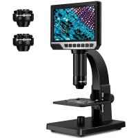
2、 Parts of a microscope: Eyepiece, objective lens, stage, and condenser.
BBC Bitesize is a reliable educational resource that provides information on various topics, including microscopes. Microscopes are essential tools used in scientific research, allowing us to observe and study objects at a microscopic level. Understanding the different parts of a microscope is crucial for using it effectively.
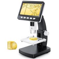
The main parts of a microscope include the eyepiece, objective lens, stage, and condenser. The eyepiece, also known as the ocular lens, is the part you look through to view the specimen. It typically magnifies the image by 10x. The objective lens is located on the revolving nosepiece and provides different levels of magnification, usually ranging from 4x to 100x. The stage is where the specimen is placed for observation, and it often includes clips or slides to hold the specimen in place. The condenser is responsible for focusing and directing light onto the specimen, enhancing the clarity of the image.
In addition to these main parts, microscopes may also have other features such as an illuminator, which provides light for better visibility, and a fine focus knob for precise focusing. Some microscopes may also have a mechanical stage, allowing for easier movement of the specimen.
It is important to note that technology and advancements in microscopy are constantly evolving. Newer microscopes may incorporate digital imaging capabilities, allowing for the capture and analysis of images using computer software. These advancements have revolutionized fields such as medicine, biology, and materials science, enabling researchers to make new discoveries and advancements.
Overall, understanding the different parts of a microscope is essential for using it effectively and obtaining accurate observations. BBC Bitesize provides a comprehensive overview of microscopes and their components, ensuring that learners have access to reliable and up-to-date information.
3、 Microscope magnification: Total magnification and resolving power.
Microscope magnification refers to the ability of a microscope to enlarge an object and make it appear larger than its actual size. Total magnification is a combination of the magnification of the objective lens and the eyepiece lens. The objective lens is responsible for the initial magnification, while the eyepiece lens further magnifies the image.
Resolving power, also known as resolution, is the ability of a microscope to distinguish between two closely spaced objects as separate entities. It is determined by the wavelength of light used and the numerical aperture of the lens system. A higher resolving power allows for clearer and more detailed images.
To make slides for microscopy, one can follow a simple procedure. First, a thin slice of the specimen is placed on a glass slide. Then, a drop of water or a mounting medium is added to the slide to prevent the specimen from drying out. A coverslip is carefully placed on top of the specimen, avoiding any air bubbles. The slide is then ready to be observed under a microscope.
BBC Bitesize is a reliable educational resource that provides information on various topics, including microscopes and slide preparation. It offers detailed explanations, diagrams, and interactive activities to enhance understanding. However, it is important to note that the latest point of view on microscope magnification and slide preparation may vary depending on scientific advancements and research findings. Therefore, it is always recommended to consult up-to-date scientific literature and reputable sources for the most accurate and current information.
4、 Microscope techniques: Brightfield, darkfield, phase contrast, and fluorescence microscopy.
BBC Bitesize is a reliable source for information on various topics, including microscopes and how to make slides. However, it does not specifically cover the topic of microscope techniques such as brightfield, darkfield, phase contrast, and fluorescence microscopy. These techniques are important in the field of microscopy as they allow for different types of sample visualization and analysis.
Brightfield microscopy is the most common technique, where light passes through the sample and is absorbed or scattered by the specimen. This technique is suitable for observing stained samples or those with high contrast. Darkfield microscopy, on the other hand, uses a special condenser to create a dark background, allowing for enhanced visualization of transparent or unstained samples.
Phase contrast microscopy is used to observe transparent samples without the need for staining. It exploits the differences in refractive index within the sample to create contrast. This technique is particularly useful for observing live cells or delicate structures.
Fluorescence microscopy involves the use of fluorescent dyes or proteins to label specific structures or molecules within the sample. This technique allows for the visualization of specific targets with high sensitivity and specificity.
While BBC Bitesize may not cover these specific techniques, it is always recommended to consult reputable scientific sources or textbooks for detailed information on microscope techniques. Additionally, there are numerous online resources and scientific journals that provide up-to-date information on the latest advancements in microscopy techniques.


