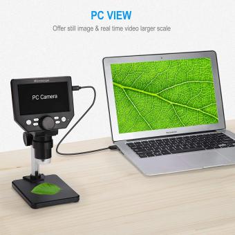
Can You See An Atom In A Microscope ?
No, atoms cannot be directly seen using a traditional light microscope due to their extremely small size. The wavelength of visible light is much larger than the size of an atom, making it impossible to resolve individual atoms using this type of microscope. However, there are specialized microscopes such as scanning tunneling microscopes and atomic force microscopes that can indirectly visualize atoms by detecting their interactions with a probe. These techniques rely on the principles of quantum mechanics and use a sharp tip to scan the surface of a sample, providing a detailed image of the atomic arrangement.
1、 Atomic Force Microscopy: Visualizing atoms using a scanning probe technique.
Can you see an atom in a microscope? The answer is yes, but not with a conventional light microscope. Atoms are much smaller than the wavelength of visible light, making them impossible to directly observe using traditional optical microscopes. However, with the advent of advanced imaging techniques such as Atomic Force Microscopy (AFM), it is now possible to visualize atoms.
AFM is a scanning probe technique that allows for the imaging of surfaces at the atomic level. It works by using a tiny probe, typically a sharp tip at the end of a cantilever, to scan the surface of a sample. As the probe moves across the surface, it experiences forces that are used to create a topographic map of the sample.
The resolution of AFM is so high that individual atoms can be observed. By scanning the probe over a sample, the tip can detect the atomic-scale features and create a detailed image of the surface. This technique has revolutionized the field of nanoscience and has provided valuable insights into the structure and behavior of materials at the atomic level.
It is important to note that AFM is not the only technique capable of visualizing atoms. Scanning Tunneling Microscopy (STM) is another powerful tool that can image individual atoms on conducting surfaces. Both AFM and STM have been instrumental in advancing our understanding of nanoscale phenomena and have opened up new possibilities for manipulating and engineering materials at the atomic level.
In conclusion, while atoms cannot be directly observed using a conventional light microscope, techniques such as Atomic Force Microscopy and Scanning Tunneling Microscopy have made it possible to visualize atoms and study their properties in great detail. These advancements have significantly contributed to our understanding of the nanoworld and have paved the way for numerous technological advancements.
2、 Scanning Tunneling Microscopy: Imaging atoms by measuring electron tunneling.
Scanning Tunneling Microscopy (STM) is a powerful technique that allows scientists to image atoms by measuring electron tunneling. With STM, it is indeed possible to see individual atoms under certain conditions.
The principle behind STM involves scanning a sharp metal tip across a sample surface while maintaining a small distance between the tip and the surface. A voltage is applied between the tip and the sample, creating a tunneling current of electrons. By monitoring this current, scientists can map the surface topography at the atomic scale.
The resolution of STM is remarkable, with the ability to visualize individual atoms and even their electronic structure. This technique has been instrumental in advancing our understanding of materials science, nanotechnology, and surface physics. It has allowed scientists to study the arrangement of atoms on surfaces, investigate the behavior of molecules, and explore the properties of various materials.
However, it is important to note that STM has its limitations. The technique requires a conductive sample surface, which restricts its application to certain materials. Additionally, STM operates under ultra-high vacuum conditions, making it challenging to study samples in real-world environments.
In recent years, advancements in microscopy techniques have expanded our ability to visualize atoms. For example, aberration-corrected transmission electron microscopy (TEM) has achieved atomic resolution imaging, allowing scientists to directly observe individual atoms in three dimensions. This technique has opened up new possibilities for studying complex materials and biological systems.
In conclusion, while STM is a powerful tool for imaging atoms, it is not the only technique available. With the continuous development of microscopy techniques, scientists now have multiple methods to visualize atoms and gain insights into the atomic world.

3、 Transmission Electron Microscopy: Resolving atomic structures using electron beams.
Transmission Electron Microscopy (TEM) is a powerful technique that allows scientists to observe and analyze the atomic structure of materials. With TEM, it is indeed possible to see individual atoms, providing valuable insights into the arrangement and behavior of matter at the atomic level.
In TEM, a beam of electrons is transmitted through a thin sample, and the resulting image is formed by the interaction of the electrons with the atoms in the sample. The resolution of TEM is much higher than that of traditional light microscopes, allowing for the visualization of atomic structures. The latest advancements in TEM technology have further improved the resolution, enabling scientists to observe individual atoms with remarkable clarity.
However, it is important to note that directly "seeing" an atom in a TEM image is not as straightforward as observing a macroscopic object. Atoms are extremely small, and their images in TEM are often represented as bright or dark spots, depending on their electron scattering properties. The interpretation of these images requires expertise and careful analysis.
Moreover, the ability to observe individual atoms in TEM is influenced by various factors, including the quality of the sample, the electron beam energy, and the imaging conditions. Achieving atomic resolution in TEM often requires specialized techniques, such as high-resolution imaging or electron diffraction.
In summary, while it is possible to see individual atoms using Transmission Electron Microscopy, it is a complex process that requires advanced techniques and expertise. The latest advancements in TEM technology have significantly improved the resolution, allowing scientists to explore and understand the atomic world with unprecedented detail.

4、 Scanning Electron Microscopy: Obtaining high-resolution images of atomic surfaces.
Can you see an atom in a microscope? The answer is no, you cannot see an individual atom using a traditional light microscope. This is because the wavelength of visible light is much larger than the size of an atom, making it impossible to resolve individual atoms using this type of microscope.
However, with the advent of advanced imaging techniques such as Scanning Electron Microscopy (SEM), it is now possible to obtain high-resolution images of atomic surfaces. SEM uses a beam of electrons instead of light to image the sample, allowing for much higher magnification and resolution.
In SEM, a focused beam of electrons is scanned across the surface of the sample, and the interaction between the electrons and the atoms in the sample produces signals that can be detected and used to create an image. By carefully controlling the parameters of the electron beam and the detectors, it is possible to obtain images with resolutions on the order of a few nanometers, which is small enough to observe individual atoms or atomic arrangements on the surface of a material.
It is important to note that while SEM can provide high-resolution images of atomic surfaces, it does not allow for direct visualization of individual atoms in three dimensions. To achieve this level of detail, more advanced techniques such as transmission electron microscopy (TEM) or scanning tunneling microscopy (STM) are required.
In conclusion, while you cannot see an individual atom using a traditional light microscope, Scanning Electron Microscopy offers a powerful tool for obtaining high-resolution images of atomic surfaces. However, it is important to use the appropriate imaging technique depending on the level of detail required.
