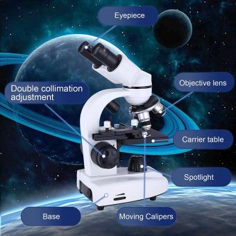
How Does An Electron Microscope Work Simple ?
An electron microscope works by using a beam of electrons instead of light to magnify objects. The electrons are focused using electromagnetic lenses and directed towards the object being studied. As the electrons interact with the object, they create an image that can be viewed on a screen or captured using a camera. The resolution of an electron microscope is much higher than that of a traditional light microscope, allowing for the visualization of much smaller structures and details.
1、 Electron beam generation

Electron microscopes work by using a beam of electrons instead of light to magnify objects. The electron beam is generated by heating a filament, similar to a light bulb, which releases electrons. These electrons are then accelerated through a series of electromagnetic lenses, which focus the beam onto the object being studied.
As the electron beam interacts with the object, it scatters and reflects off the surface, creating a signal that is detected by a detector. This signal is then processed and used to create an image of the object.
One of the key advantages of electron microscopes is their ability to achieve much higher magnification than traditional light microscopes. This is because electrons have a much shorter wavelength than visible light, allowing them to resolve much smaller details.
In recent years, advances in electron microscopy have led to the development of new techniques such as cryo-electron microscopy, which allows researchers to study biological samples in their native state. This has led to a better understanding of the structure and function of proteins and other biomolecules, and has the potential to revolutionize drug discovery and development.
Overall, electron microscopes are powerful tools for studying the structure and properties of materials at the nanoscale, and continue to play an important role in scientific research and discovery.
2、 Electromagnetic lenses

An electron microscope works by using a beam of electrons instead of light to magnify objects. The electrons are accelerated through a vacuum and focused onto the object being studied using electromagnetic lenses. These lenses are similar to the lenses in a traditional microscope, but they use magnetic fields to bend and focus the electron beam instead of glass lenses.
As the electrons interact with the object, they scatter and produce signals that can be detected and used to create an image. The image is then displayed on a screen or captured digitally for further analysis.
One of the advantages of electron microscopes is their ability to achieve much higher magnification than traditional light microscopes. This allows researchers to study objects at the atomic and molecular level, providing insights into the structure and behavior of materials and biological specimens.
Recent advances in electron microscopy have also led to the development of new techniques, such as cryo-electron microscopy, which allows researchers to study biological samples in their natural state without the need for staining or fixation. This has opened up new avenues for understanding the structure and function of complex biological systems.
Overall, electron microscopes are powerful tools for scientific research and have contributed to many important discoveries in fields ranging from materials science to biology.
3、 Sample preparation

How does an electron microscope work simple? An electron microscope works by using a beam of electrons instead of light to magnify objects. The electrons are focused using electromagnetic lenses and directed onto the sample, which is usually very thin and coated with a conductive material to prevent charging. As the electrons interact with the sample, they scatter and produce an image that is detected by a sensor and displayed on a screen.
Sample preparation is a crucial step in electron microscopy. Samples must be thin enough to allow electrons to pass through and produce a clear image. This can be achieved through techniques such as slicing, grinding, or chemical etching. In addition, samples must be coated with a conductive material to prevent charging, which can distort the image.
Recent advances in electron microscopy have allowed for even higher resolution imaging. Cryo-electron microscopy, for example, allows samples to be imaged at near-atomic resolution by freezing them in a thin layer of ice. This technique has revolutionized the study of biological molecules and has led to new insights into the structure and function of proteins and other biomolecules.
In summary, electron microscopes work by using a beam of electrons to magnify objects, and sample preparation is a critical step in achieving high-quality images. Advances in electron microscopy continue to push the boundaries of what is possible, allowing for ever more detailed and precise imaging of the microscopic world.
4、 Scanning vs. transmission electron microscopy

How does an electron microscope work simply?
An electron microscope works by using a beam of electrons instead of light to magnify objects. The electrons are focused using electromagnetic lenses and directed onto the sample, which is placed in a vacuum to prevent interference from air molecules. The electrons interact with the sample, producing signals that are detected and used to create an image.
There are two main types of electron microscopy: scanning electron microscopy (SEM) and transmission electron microscopy (TEM). SEM produces images by scanning the surface of the sample with a focused beam of electrons, while TEM passes the beam through the sample to produce an image of the internal structure.
In recent years, advancements in electron microscopy technology have allowed for even higher resolution imaging, including the ability to capture images of individual atoms. This has led to new insights in fields such as materials science, biology, and nanotechnology.
Overall, electron microscopy has become an essential tool for researchers in a wide range of fields, allowing them to see and understand the world at a level of detail that was previously impossible.


