How Does An Endoscope Work Total Internal Reflection ?
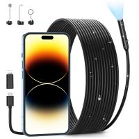
An endoscope is a medical device that is used to examine the internal organs and cavities of the human body. It consists of a long, thin, flexible tube with a camera and light source at the end. The camera captures images of the internal organs and sends them to a monitor for the doctor to view.
Total internal reflection is a principle of optics that is used in endoscopes to transmit light through the tube. The light source at the end of the endoscope emits light, which is then reflected off the internal walls of the tube. The walls of the tube are made of a material that has a higher refractive index than the surrounding air, which causes the light to be reflected back into the tube.
As the light travels through the tube, it is focused by a lens onto the internal organs. The camera at the end of the tube captures the reflected light and sends it to a monitor for the doctor to view. The use of total internal reflection allows the endoscope to transmit light through the tube without losing much of its intensity, which is important for obtaining clear images of the internal organs.
1、 Optical Fiber
An endoscope is a medical device used to examine the internal organs and cavities of the human body. It consists of a long, thin, flexible tube with a camera and light source at the end. The camera captures images of the internal organs and sends them to a monitor for viewing by the doctor.
The endoscope uses optical fibers to transmit light from the light source to the tip of the tube. Optical fibers are made of glass or plastic and are designed to transmit light through total internal reflection. Total internal reflection occurs when light is reflected back into the fiber instead of being refracted out of it. This allows the light to travel long distances without losing intensity or clarity.
The light at the tip of the endoscope illuminates the internal organs, allowing the camera to capture clear images. The camera then sends the images back through the optical fibers to the monitor for viewing.
The use of optical fibers in endoscopes has revolutionized medical imaging. It allows doctors to see inside the body without the need for invasive surgery. The latest advancements in endoscope technology include high-definition cameras and 3D imaging, which provide even clearer and more detailed images.
In conclusion, the endoscope works by using optical fibers to transmit light from the light source to the tip of the tube. Total internal reflection allows the light to travel long distances without losing intensity or clarity, making it possible for doctors to see inside the body without invasive surgery. The latest advancements in endoscope technology continue to improve the quality and accuracy of medical imaging.
2、 Light Source
An endoscope is a medical device used to examine the internal organs and cavities of the human body. It consists of a long, thin, flexible tube with a camera and light source at the end. The light source is an essential component of the endoscope, as it illuminates the area being examined, allowing the camera to capture clear images.
The light source in an endoscope works on the principle of total internal reflection. Total internal reflection occurs when light travels from a medium with a higher refractive index to a medium with a lower refractive index. When this happens, the light is reflected back into the higher refractive index medium, rather than passing through the lower refractive index medium.
In an endoscope, the light source is typically a fiber optic cable. The cable is made up of thousands of tiny fibers, each with a core of high refractive index glass surrounded by a cladding of lower refractive index glass. When light is introduced into one end of the cable, it travels down the core of each fiber through total internal reflection until it reaches the other end of the cable.
The latest point of view on endoscope technology is the development of miniaturized endoscopes that can be swallowed like a pill. These endoscopes use wireless technology to transmit images to an external receiver, eliminating the need for a long, flexible tube. They are less invasive and more comfortable for patients, making them a promising technology for the future of medical imaging.
3、 Lens System
An endoscope is a medical device used to examine the internal organs and cavities of the human body. It consists of a long, thin, flexible tube with a camera and light source at one end. The camera captures images of the internal organs and sends them to a monitor for the doctor to view.
One of the key components of an endoscope is the lens system. The lens system is responsible for capturing high-quality images of the internal organs. It consists of a series of lenses that work together to focus the light from the light source onto the camera sensor.
One of the key principles behind the functioning of the lens system in an endoscope is total internal reflection. Total internal reflection occurs when light travels from a medium with a higher refractive index to a medium with a lower refractive index. When this happens, the light is reflected back into the higher refractive index medium, rather than passing through the lower refractive index medium.
In an endoscope, the lens system is designed to use total internal reflection to capture high-quality images of the internal organs. The lenses are made from materials with a high refractive index, such as glass or plastic. When light enters the lens, it is refracted and focused onto the camera sensor. The use of total internal reflection ensures that the light is not lost as it passes through the lens, resulting in a clear and sharp image.
In recent years, there have been advancements in endoscope technology that have improved the functioning of the lens system. For example, some endoscopes now use digital sensors instead of traditional cameras, resulting in higher resolution images. Additionally, some endoscopes now use advanced imaging techniques, such as fluorescence imaging, to improve the detection of abnormalities in the internal organs.

4、 CCD Camera
An endoscope is a medical device used to examine the internal organs and cavities of the human body. It consists of a long, thin, flexible tube with a camera and light source at the end. The camera captures images of the internal organs and sends them to a monitor for the doctor to view.
One of the key components of an endoscope is the CCD (charge-coupled device) camera. The CCD camera is a type of digital camera that converts light into electrical signals. It is located at the tip of the endoscope and captures high-quality images of the internal organs.
The CCD camera works by using a process called total internal reflection. Total internal reflection occurs when light travels from a denser medium, such as glass or water, to a less dense medium, such as air. When this happens, the light is reflected back into the denser medium instead of passing through the less dense medium.
In an endoscope, the light source at the tip of the device shines light onto the internal organs. The light then reflects off the organs and enters the endoscope tube. The tube is lined with a reflective material that reflects the light towards the CCD camera. The camera then captures the reflected light and converts it into an electrical signal that is sent to the monitor.
The latest point of view on endoscopes is the development of wireless endoscopes. These devices use wireless technology to transmit images from the camera to a monitor, eliminating the need for a physical cable. This makes the endoscope more flexible and easier to use, as it can be moved around more freely. Additionally, wireless endoscopes are less invasive and more comfortable for the patient.


