How Does The Endoscope Work ?
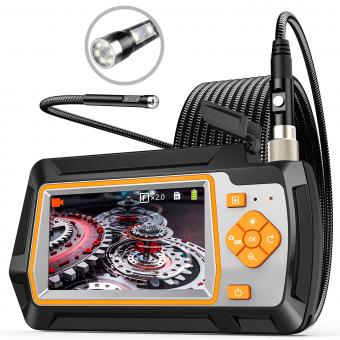
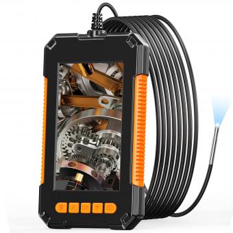
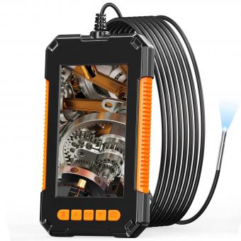
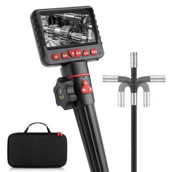
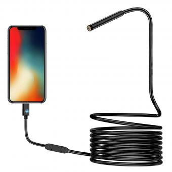
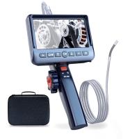
An endoscope is a medical device that is used to examine the inside of the body. It consists of a long, thin, flexible tube with a light and a camera at the end. The endoscope is inserted into the body through a natural opening or a small incision, and the camera sends images to a monitor, allowing the doctor to see inside the body.
The endoscope can be used to examine various parts of the body, such as the digestive system, respiratory system, urinary system, and reproductive system. It can also be used to perform procedures such as biopsies, removing foreign objects, and treating bleeding.
The endoscope works by using a series of lenses and mirrors to transmit light and images from the camera at the end of the tube to the monitor. The tube is flexible, allowing it to be maneuvered through the body's curves and bends. The light source illuminates the area being examined, and the camera captures images that are transmitted to the monitor in real-time. This allows the doctor to see any abnormalities or issues that may be present and make a diagnosis or perform a procedure as needed.
1、 Optical System

How does the endoscope work? The answer lies in its optical system. An endoscope is a medical device that allows doctors to see inside the body without making large incisions. The optical system of an endoscope consists of a lens, a light source, and a camera. The lens is used to focus the light onto the area being examined, while the camera captures the images and sends them to a monitor for the doctor to view.
The latest advancements in endoscope technology have led to the development of high-definition cameras and improved lighting systems. These advancements have greatly improved the quality of images captured by endoscopes, making it easier for doctors to diagnose and treat medical conditions.
In addition to medical applications, endoscopes are also used in other fields such as industrial inspection and veterinary medicine. The use of endoscopes in these fields has also benefited from advancements in optical technology.
Overall, the optical system of an endoscope plays a crucial role in its functionality. The ability to capture high-quality images is essential for accurate diagnosis and treatment of medical conditions. As technology continues to advance, we can expect further improvements in endoscope optical systems, leading to even better medical outcomes.
2、 Light Source
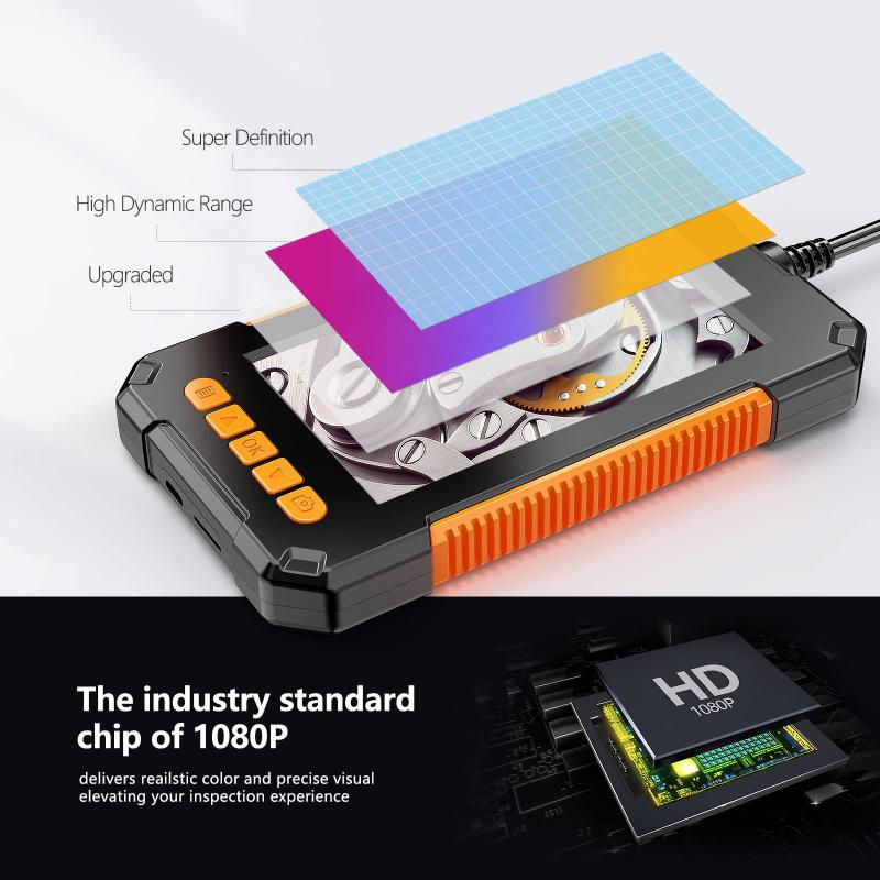
How does the endoscope work? One of the key components of an endoscope is the light source. The light source provides illumination to the area being examined, allowing the endoscope's camera to capture clear images. The light source is typically an LED or fiber optic cable that is connected to the endoscope's camera head.
The latest advancements in endoscope technology have led to the development of high-definition cameras and improved lighting systems. These advancements have greatly improved the quality of images captured by endoscopes, making it easier for doctors to diagnose and treat medical conditions.
In addition to the light source, endoscopes also have a flexible tube that can be inserted into the body. The tube is typically made of a flexible material such as rubber or plastic, and it can be manipulated by the doctor to navigate through the body's internal organs.
Once the endoscope is inserted into the body, the camera captures images that are transmitted to a monitor. The doctor can then use these images to examine the area being examined and make a diagnosis.
Overall, the endoscope is a valuable tool in modern medicine, allowing doctors to examine internal organs and diagnose medical conditions without the need for invasive surgery. The light source is just one of the many components that make this technology possible, and ongoing advancements in endoscope technology are sure to lead to even more improvements in the years to come.
3、 Fiber Optics
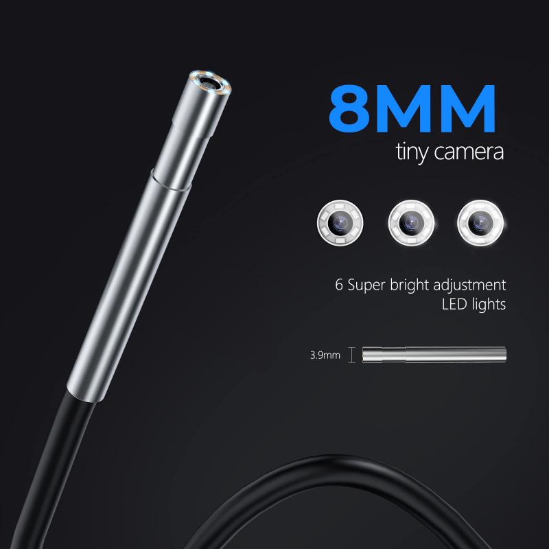
How does the endoscope work? The answer lies in the use of fiber optics. Fiber optics is a technology that uses thin, flexible glass or plastic fibers to transmit light signals. In an endoscope, a bundle of these fibers is used to transmit light from a source to the area being examined. The light reflects off the tissue and is transmitted back through the fibers to a camera, which captures the image and displays it on a monitor.
The use of fiber optics in endoscopes has revolutionized medical imaging. It allows doctors to see inside the body without making large incisions, reducing the risk of complications and speeding up recovery times. Endoscopes are used to examine the digestive tract, respiratory system, urinary tract, and other areas of the body.
Recent advancements in fiber optics technology have further improved the capabilities of endoscopes. For example, high-definition cameras and improved lighting systems provide clearer images, allowing doctors to make more accurate diagnoses. Additionally, the use of miniature cameras and sensors has enabled the development of capsule endoscopes, which can be swallowed and travel through the digestive tract, providing a non-invasive way to examine the small intestine.
In conclusion, the use of fiber optics in endoscopes has revolutionized medical imaging and allowed doctors to see inside the body without making large incisions. Recent advancements in fiber optics technology have further improved the capabilities of endoscopes, providing clearer images and enabling the development of non-invasive capsule endoscopes.
4、 Camera and Image Processing

How does the endoscope work? The endoscope is a medical device that allows doctors to see inside the body without making large incisions. It consists of a long, thin tube with a camera and light source at the end. The camera captures images of the inside of the body and sends them to a monitor for the doctor to view.
The camera and light source work together to illuminate the area being examined. The light source is usually an LED or fiber optic cable that transmits light to the end of the tube. The camera captures the reflected light and converts it into an image that can be viewed on a monitor.
Image processing is also an important part of how the endoscope works. The images captured by the camera are often distorted or blurry due to the curvature of the tube and the movement of the body. Image processing software is used to correct these distortions and enhance the image quality.
The latest advancements in endoscope technology include the use of high-definition cameras and 3D imaging. These advancements allow doctors to see even more detail and improve their ability to diagnose and treat medical conditions.
In conclusion, the endoscope works by using a camera and light source to capture images of the inside of the body. Image processing software is used to correct distortions and enhance image quality. The latest advancements in endoscope technology have improved the ability of doctors to diagnose and treat medical conditions.


