How Phase Contrast Microscope Works?
A phase contrast microscope works by utilizing the phase shifts of light passing through a transparent specimen to create contrast in the image. This is achieved by using a special condenser and objective lens that manipulate the phase of light waves passing through the specimen. The phase shifts caused by the specimen are then converted into differences in brightness, creating contrast in the image without the need for staining or labeling the sample. This technique is particularly useful for observing live cells and other transparent specimens in their natural state.
1、 Principle of Phase Contrast Microscopy

The principle of phase contrast microscopy is based on the fact that light waves passing through different materials can undergo a phase shift, which is a change in the position of the wave relative to a reference point. In a phase contrast microscope, this phase shift is utilized to enhance the contrast of transparent and colorless specimens, such as living cells, which would otherwise be difficult to visualize using traditional brightfield microscopy.
The phase contrast microscope achieves this by using a special condenser and objective lens that manipulate the phase of the light passing through the specimen. The condenser creates a hollow cone of light that illuminates the specimen, and the phase shifts caused by the specimen are then converted into differences in brightness, creating contrast in the image. This allows for the visualization of internal structures and details within the specimen that would be otherwise invisible.
In recent years, advancements in phase contrast microscopy have included the development of digital phase contrast techniques, which utilize digital image processing to further enhance the contrast and resolution of phase contrast images. Additionally, the integration of phase contrast microscopy with other imaging modalities, such as fluorescence microscopy, has allowed for the simultaneous visualization of both structural and functional information within biological specimens.
Overall, the principle of phase contrast microscopy continues to be a valuable tool in biological research, enabling the visualization of dynamic processes and delicate structures within living cells.
2、 Components of Phase Contrast Microscope
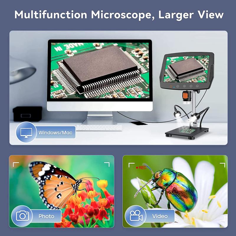
The phase contrast microscope works by taking advantage of the phase differences in light waves that pass through different parts of a specimen. This technique allows for the visualization of transparent or colorless samples that would otherwise be difficult to see with a standard bright-field microscope. The key components of a phase contrast microscope include a condenser, phase plate, objective lens, and eyepiece.
The condenser focuses light onto the specimen, and the phase plate, which is located in the condenser, shifts the phase of the light waves passing through the specimen. As the light waves pass through the specimen, they are either slowed down or sped up, creating phase differences. The objective lens then collects the light and focuses it onto the eyepiece, where the phase differences are converted into differences in brightness, allowing for the visualization of the specimen.
The latest point of view on phase contrast microscopy emphasizes the importance of digital imaging and analysis. Advanced digital cameras and software allow for the capture and analysis of phase contrast images, enabling researchers to quantify and measure phase differences in specimens with high precision. This has expanded the applications of phase contrast microscopy in fields such as cell biology, microbiology, and materials science. Additionally, advancements in optical technology have led to the development of high-performance phase contrast microscope systems with improved resolution and sensitivity, further enhancing the capabilities of this technique.
3、 Working of Phase Plate in Phase Contrast Microscope

The phase contrast microscope works by utilizing the phase differences of light passing through different parts of a specimen to create contrast in the image. This is achieved through the use of a phase plate, which is a specialized optical component that introduces a phase shift to the light passing through the specimen. The phase plate consists of a transparent material with a specific thickness and refractive index, which alters the phase of the light passing through it.
When light passes through the specimen, it interacts with the different structures and components, causing variations in the phase of the light waves. These phase differences are typically too subtle to be detected by a standard brightfield microscope, resulting in low contrast images. However, the phase plate in a phase contrast microscope introduces a phase shift to the light passing through the specimen, converting the phase differences into intensity differences that can be visualized.
The latest point of view on the working of the phase plate in a phase contrast microscope emphasizes the importance of precise engineering and design of the phase plate to achieve optimal contrast and resolution. Advanced computational techniques and machine learning algorithms are also being employed to enhance the image processing and analysis capabilities of phase contrast microscopy, allowing for more detailed and accurate visualization of biological specimens.
In summary, the phase plate in a phase contrast microscope plays a crucial role in converting phase differences in light passing through a specimen into visible contrast, enabling the visualization of transparent and low contrast biological samples with high resolution and clarity.
4、 Advantages of Phase Contrast Microscopy

Phase contrast microscopy is a technique used to enhance the contrast of transparent, unstained samples, such as living cells, by exploiting the phase shifts that occur when light passes through different parts of the sample. This is achieved by using a special condenser and objective lens that manipulate the phase of the light passing through the sample, resulting in the conversion of phase shifts into changes in brightness, which can be visualized as contrast in the image.
The phase contrast microscope works by splitting the light into two paths, one passing through the sample and the other passing through a phase plate. The phase plate introduces a phase shift to the light passing through it, which is then combined with the light passing through the sample. This results in interference patterns that are converted into contrast in the final image.
The advantages of phase contrast microscopy include the ability to visualize transparent samples without the need for staining, which can alter the sample's natural properties. It also allows for the observation of living cells in real time, providing valuable insights into dynamic biological processes. Additionally, phase contrast microscopy is a non-destructive technique, making it suitable for long-term observation of samples.
From the latest point of view, phase contrast microscopy has become increasingly important in fields such as cell biology, microbiology, and medical research, as it enables the visualization of delicate structures and processes within living cells. It has also found applications in the study of neurobiology and developmental biology, where the ability to observe dynamic processes in real time is crucial. Furthermore, advancements in phase contrast microscopy technology have led to improved image quality and resolution, making it an indispensable tool for modern biological research.


