How To Measure Something Under A Microscope ?
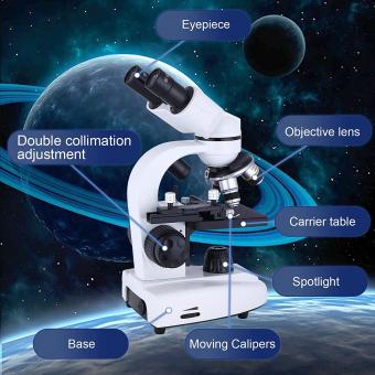
To measure something under a microscope, you can use an eyepiece reticle or a stage micrometer. An eyepiece reticle is a small glass disk with a scale etched onto it that fits into the eyepiece of the microscope. By focusing on the object and aligning it with the scale, you can measure its size. A stage micrometer is a glass slide with a precise scale etched onto it that is placed on the microscope stage. By comparing the scale on the stage micrometer to the scale on the eyepiece reticle, you can determine the size of the object being viewed. Additionally, some microscopes have built-in measurement software that allows you to measure the size of objects directly on the computer screen.
1、 Magnification and Resolution
How to measure something under a microscope is a common question among scientists and researchers. The answer lies in understanding the concepts of magnification and resolution. Magnification refers to the degree to which an object is enlarged under the microscope, while resolution refers to the ability to distinguish between two closely spaced objects.
To measure something under a microscope, one must first select the appropriate magnification level. This will depend on the size of the object being observed and the level of detail required. Once the object is in focus, measurements can be taken using a calibrated eyepiece or a stage micrometer.
However, it is important to note that the resolution of the microscope also plays a crucial role in accurate measurements. The higher the resolution, the more detail can be observed and measured. Advances in technology have led to the development of high-resolution microscopes, such as electron microscopes, which can provide unprecedented levels of detail.
In addition to magnification and resolution, other factors such as lighting and contrast can also affect the accuracy of measurements taken under a microscope. Proper sample preparation and handling are also important to ensure accurate measurements.
In conclusion, measuring something under a microscope requires an understanding of magnification and resolution, as well as attention to other factors that can affect accuracy. With the latest advances in technology, researchers can now obtain highly detailed measurements that were once impossible.
2、 Field of View
How to measure something under a microscope? One of the most important concepts to understand when measuring objects under a microscope is the field of view. The field of view refers to the area that is visible through the microscope lens at any given time. It is typically measured in millimeters or micrometers and can vary depending on the magnification of the lens being used.
To measure an object under a microscope, you first need to determine the field of view. This can be done by placing a ruler or micrometer slide under the microscope and measuring the distance across the field of view. Once you know the field of view, you can then measure the object by placing it under the microscope and using the ruler or micrometer slide to measure its size in relation to the field of view.
It is important to note that the field of view can change depending on the magnification of the lens being used. As the magnification increases, the field of view decreases, making it more difficult to measure objects accurately. To compensate for this, it is important to use a calibrated micrometer slide or stage micrometer to ensure accurate measurements.
In recent years, advances in technology have made it possible to measure objects under a microscope using digital imaging software. This software allows for precise measurements to be taken and can be particularly useful when measuring objects that are difficult to see or measure manually.
In conclusion, measuring objects under a microscope requires an understanding of the field of view and the use of calibrated measuring tools. With the latest advances in technology, digital imaging software can also be used to take precise measurements.
3、 Depth of Field
How to measure something under a microscope:
To measure something under a microscope, you need to first calibrate the microscope using a stage micrometer. This will allow you to determine the magnification of the microscope and the size of the field of view. Once you have calibrated the microscope, you can use it to measure the size of objects under the microscope.
One important factor to consider when measuring something under a microscope is the depth of field. The depth of field is the distance between the nearest and farthest objects in focus in a microscope image. It is important to understand the depth of field because it can affect the accuracy of your measurements.
To measure the depth of field, you can use a technique called focus stacking. This involves taking multiple images of the same object at different focal planes and then combining them into a single image using software. By doing this, you can increase the depth of field and get a more accurate measurement of the object.
Another important consideration when measuring something under a microscope is the resolution of the microscope. The resolution is the ability of the microscope to distinguish between two closely spaced objects. The higher the resolution, the more accurate your measurements will be.
In recent years, advances in technology have led to the development of new microscopy techniques, such as super-resolution microscopy, which can provide even higher resolution and more accurate measurements. These techniques are becoming increasingly important in fields such as biology and materials science, where precise measurements are essential for understanding the properties of materials and biological systems.

4、 Illumination
How to measure something under a microscope? One of the most important factors to consider is illumination. Proper illumination is crucial for obtaining clear and accurate measurements of microscopic specimens. Illumination can affect the contrast, resolution, and depth of field of the image, which in turn can affect the accuracy of the measurements.
To ensure proper illumination, it is important to use the appropriate type of microscope and lighting source. For example, a compound microscope with a brightfield illumination source is ideal for observing transparent or translucent specimens, while a darkfield illumination source is better for observing opaque specimens. Additionally, the intensity and angle of the lighting source should be adjusted to optimize the contrast and resolution of the image.
Another important consideration is the use of filters. Filters can be used to enhance contrast, reduce glare, and improve color accuracy. For example, a polarizing filter can be used to reduce glare and improve contrast, while a color filter can be used to enhance color accuracy.
In recent years, advances in technology have led to the development of new illumination techniques, such as fluorescence microscopy and confocal microscopy. These techniques use specialized lighting sources and filters to selectively illuminate specific parts of the specimen, allowing for more detailed and accurate measurements.
In conclusion, proper illumination is essential for obtaining clear and accurate measurements of microscopic specimens. By using the appropriate microscope, lighting source, and filters, and taking advantage of the latest illumination techniques, researchers can obtain high-quality images and measurements that can help advance our understanding of the microscopic world.













There are no comments for this blog.