How To See Bacteria Under A Microscope ?
To see bacteria under a microscope, you will need to follow these steps:
1. Prepare a bacterial sample: Collect a sample of bacteria from a source such as water, soil, or a culture. Ensure that the sample is pure and free from contaminants.
2. Prepare a slide: Take a clean microscope slide and place a drop of the bacterial sample onto it. Spread the drop evenly across the slide using a sterile loop or a pipette.
3. Stain the bacteria (optional): Staining the bacteria can enhance their visibility. Use a suitable stain, such as crystal violet or methylene blue, and apply it to the bacterial sample. Follow the staining instructions carefully.
4. Cover the slide: Place a coverslip over the bacterial sample to prevent it from drying out and to protect the microscope lens.
5. Set up the microscope: Turn on the microscope and adjust the light intensity. Start with the lowest magnification objective lens (usually 10x) and focus on the sample using the coarse and fine focus knobs.
6. Observe the bacteria: Once the sample is in focus, move to higher magnification objective lenses (40x or 100x) to observe the bacteria in more detail. Use the microscope's stage controls to navigate the slide and locate areas with bacteria.
7. Record your observations: Take notes or capture images of the bacteria using a camera attached to the microscope, if available.
Remember to handle the microscope and slides carefully, and always follow proper laboratory safety protocols.
1、 Microscope Preparation: Staining Techniques for Bacteria Observation
Microscope Preparation: Staining Techniques for Bacteria Observation
To see bacteria under a microscope, proper preparation and staining techniques are essential. Staining helps to enhance the visibility of bacteria by adding color to their structures, making them easier to observe and identify. Here is a step-by-step guide on how to prepare bacteria samples for microscopic observation:
1. Sample Collection: Collect a sample from the source where bacteria are suspected to be present. This could be a swab from a surface, a liquid sample, or a biological sample.
2. Smear Preparation: Take a clean glass slide and place a small drop of the sample onto it. Using a sterile loop or a cotton swab, spread the sample evenly across the slide to create a thin smear.
3. Fixation: Allow the smear to air dry completely. Once dry, heat-fix the slide by passing it through a flame a few times. This process kills the bacteria, attaches them to the slide, and prevents them from washing away during subsequent staining steps.
4. Staining: There are various staining techniques available, but the most commonly used are Gram staining and acid-fast staining. Gram staining differentiates bacteria into two major groups - Gram-positive and Gram-negative, based on their cell wall composition. Acid-fast staining is used specifically for Mycobacterium species, which have a unique cell wall structure.
5. Microscopic Observation: Once the staining process is complete, place the slide on the microscope stage and focus on the stained bacteria using the appropriate magnification. Adjust the lighting and focus until the bacteria come into clear view.
It is important to note that while staining techniques have been widely used for bacterial observation, recent advancements in microscopy, such as fluorescence microscopy and confocal microscopy, have provided new ways to visualize bacteria. These techniques allow for the use of fluorescent dyes or antibodies that specifically bind to bacterial structures, providing more detailed and specific information about the bacteria being observed.
In conclusion, proper staining techniques are crucial for observing bacteria under a microscope. By following the steps outlined above, one can prepare bacteria samples for microscopic observation and gain valuable insights into their morphology and characteristics.
2、 Microscope Settings: Adjusting Magnification and Focus for Bacteria Viewing
Microscope Settings: Adjusting Magnification and Focus for Bacteria Viewing
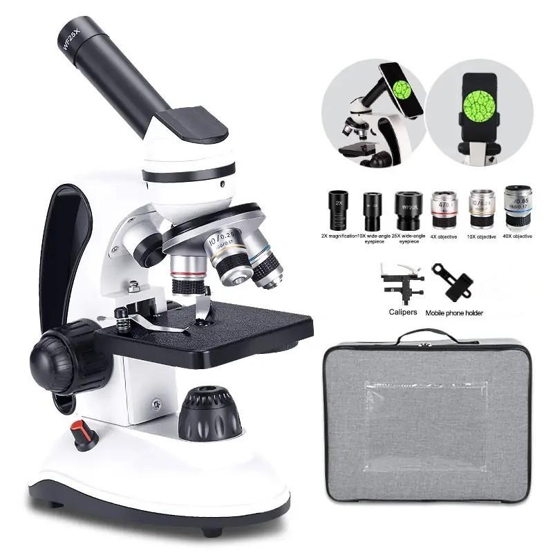
To see bacteria under a microscope, it is essential to properly adjust the magnification and focus settings. Here is a step-by-step guide on how to achieve clear and accurate observations of bacteria:
1. Prepare a bacterial sample: Start by obtaining a sample of bacteria. This can be done by swabbing a surface or collecting a liquid sample. Ensure that the sample is properly prepared, such as by spreading it on a microscope slide and allowing it to air dry.
2. Place the slide on the microscope stage: Carefully place the prepared slide on the stage of the microscope. Secure it in place using the stage clips or any other appropriate method.
3. Start with low magnification: Begin by using the lowest magnification objective lens (usually 4x or 10x) to locate the bacteria. This will provide a wider field of view, making it easier to find the bacteria.
4. Adjust the focus: Use the coarse focus knob to bring the bacteria into rough focus. Slowly turn the knob until the bacteria come into view. Once the bacteria are visible, use the fine focus knob to achieve a clear and sharp image.
5. Increase magnification: Once the bacteria are in focus, switch to a higher magnification objective lens (such as 40x or 100x) to observe the bacteria in more detail. Repeat the focus adjustment process using the coarse and fine focus knobs.
6. Use immersion oil (if necessary): For higher magnifications (typically 100x), immersion oil may be required to improve resolution. Apply a small drop of immersion oil on the slide, and carefully rotate the lens turret to bring the oil immersion lens into position.
7. Observe and record: Once the bacteria are in focus at the desired magnification, take the time to observe and record any relevant details. This could include the shape, size, and arrangement of the bacteria.
It is important to note that the latest advancements in microscopy techniques, such as fluorescence microscopy and confocal microscopy, have greatly enhanced our ability to visualize bacteria. These techniques allow for specific labeling of bacterial components or the use of fluorescent dyes to highlight bacteria in complex samples. Additionally, digital imaging and image analysis software have made it easier to capture and analyze bacterial images.
In conclusion, adjusting the magnification and focus settings on a microscope is crucial for viewing bacteria. By following the steps outlined above, one can obtain clear and accurate observations of bacteria, aiding in various scientific and medical research endeavors.

3、 Sample Preparation: Culturing and Preparing Bacterial Specimens for Microscopy
To see bacteria under a microscope, it is necessary to follow a proper sample preparation technique. One common method is culturing and preparing bacterial specimens for microscopy. Here is a step-by-step guide on how to do it:
1. Obtain a bacterial culture: Start by obtaining a pure culture of the bacteria you wish to observe. This can be obtained from a laboratory or by growing the bacteria on a suitable growth medium.
2. Prepare a slide: Take a clean glass microscope slide and sterilize it by passing it through a flame. Allow it to cool before proceeding.
3. Transfer the bacteria: Using a sterile inoculating loop or needle, transfer a small amount of the bacterial culture onto the slide. Spread the culture evenly over the slide to create a thin film.
4. Fixation: To prevent the bacteria from moving or being washed away during the staining process, the slide can be heat-fixed. Pass the slide through a flame a few times, ensuring that the bacteria are not overheated.
5. Staining: Staining the bacteria helps to enhance their visibility under the microscope. There are various staining techniques available, such as Gram staining or acid-fast staining, depending on the type of bacteria being observed. Follow the specific staining protocol accordingly.
6. Mounting: Once the staining is complete, place a coverslip over the stained bacteria. Gently press down to remove any air bubbles.
7. Microscopy: Finally, place the prepared slide under a microscope and adjust the focus to observe the bacteria. Start with a low magnification objective and gradually increase the magnification to get a closer look at the bacteria.
It is important to note that the latest point of view emphasizes the use of advanced microscopy techniques, such as fluorescence microscopy or confocal microscopy, to visualize bacteria. These techniques allow for better resolution and the ability to observe specific cellular components or processes within the bacteria. Additionally, newer methods like live-cell imaging or super-resolution microscopy provide insights into the dynamic behavior of bacteria in real-time. These advancements have greatly contributed to our understanding of bacterial structure, function, and interactions with their environment.

4、 Slide Preparation: Mounting Bacterial Samples on Microscope Slides
To see bacteria under a microscope, you need to prepare a slide with a bacterial sample. Here is a step-by-step guide on slide preparation for mounting bacterial samples on microscope slides:
1. Start by sterilizing the microscope slides and coverslips. This can be done by washing them with soap and water, followed by rinsing with distilled water, and then autoclaving or flaming them to kill any bacteria present.
2. Obtain a bacterial culture. This can be done by swabbing a surface with a sterile cotton swab or by using a liquid culture obtained from a laboratory.
3. Place a small drop of the bacterial culture onto the center of a clean microscope slide.
4. Using a sterile loop or pipette, spread the bacterial culture evenly across the slide, creating a thin film. Be careful not to press too hard, as this can damage the bacteria.
5. Allow the slide to air dry completely. This can take anywhere from a few minutes to an hour, depending on the size of the bacterial sample.
6. Once the slide is dry, heat-fix the bacteria by passing the slide through a flame a few times. This helps to kill the bacteria and adhere them to the slide.
7. Stain the bacterial sample if desired. Common stains used for bacterial samples include Gram stain, which differentiates bacteria into Gram-positive and Gram-negative, and methylene blue, which provides contrast.
8. Place a coverslip over the stained bacterial sample. Gently press down to remove any air bubbles.
9. The slide is now ready to be observed under a microscope. Place the slide on the microscope stage and adjust the focus to see the bacteria clearly.
It is important to note that the above steps are general guidelines for slide preparation. Depending on the specific bacteria being studied, additional steps or modifications may be required. Additionally, it is crucial to follow proper safety protocols and handle bacterial samples in a controlled environment to prevent contamination and ensure accurate observations.
In recent years, advancements in microscopy techniques have allowed for more detailed and precise visualization of bacteria. Techniques such as fluorescence microscopy, confocal microscopy, and electron microscopy have provided researchers with the ability to study bacterial structures, interactions, and behaviors at a higher resolution. These techniques often require specialized equipment and expertise, but they offer valuable insights into the world of bacteria.














There are no comments for this blog.