Microscope How It Works ?
A microscope works by using lenses to magnify small objects or details that are not visible to the naked eye. It typically consists of an objective lens, an eyepiece, and a light source. The objective lens collects light from the specimen and forms an enlarged image. This image is then further magnified by the eyepiece, which allows the viewer to see the details. The light source illuminates the specimen, making it easier to observe. Microscopes can be either light microscopes, which use visible light, or electron microscopes, which use a beam of electrons to create the image.
1、 Optical Components and Light Path in Microscopes
A microscope is an instrument used to magnify small objects that are not visible to the naked eye. It works by utilizing a combination of optical components and a specific light path to enhance the resolution and clarity of the image.
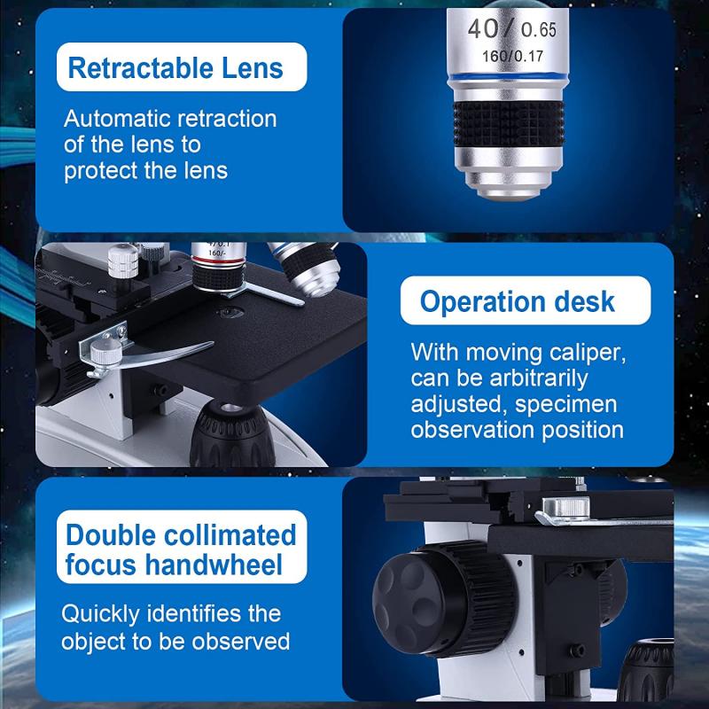
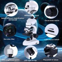
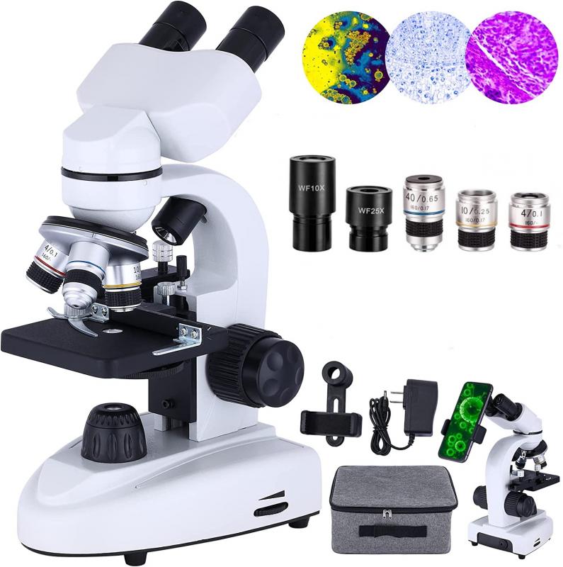
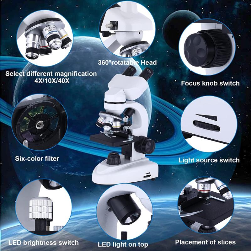
The basic components of a microscope include an objective lens, an eyepiece, and a light source. The objective lens is responsible for gathering light from the specimen and forming a magnified image. It is typically composed of multiple lenses that work together to minimize aberrations and improve image quality. The eyepiece, on the other hand, further magnifies the image formed by the objective lens and allows the viewer to observe it.
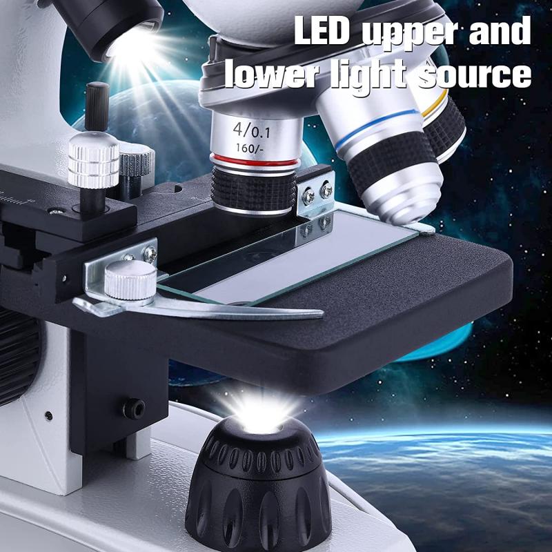
The light path in a microscope is designed to ensure that the light passes through the specimen and reaches the objective lens. The light source, which can be a lamp or an LED, provides illumination that is directed towards the specimen. The light then passes through a condenser, which focuses and concentrates the light onto the specimen. This helps to enhance the contrast and visibility of the specimen.
In recent years, there have been advancements in microscope technology, particularly in the field of microscopy. For example, the development of confocal microscopy has allowed for the imaging of three-dimensional structures with high resolution. Additionally, the use of fluorescence microscopy has enabled the visualization of specific molecules or structures within a specimen by labeling them with fluorescent dyes.
Overall, the optical components and light path in microscopes play a crucial role in enhancing the resolution and clarity of the images obtained. With advancements in technology, microscopes continue to evolve, allowing scientists and researchers to explore the microscopic world with greater detail and precision.
2、 Principles of Magnification and Resolution in Microscopy
A microscope is an instrument used to magnify and observe small objects that are not visible to the naked eye. It works by utilizing the principles of magnification and resolution.
Magnification is the process of enlarging an object to make it appear larger than its actual size. In a microscope, this is achieved through the use of lenses. The two main types of lenses used in microscopes are the objective lens and the eyepiece lens. The objective lens is located near the specimen and provides the initial magnification, while the eyepiece lens further magnifies the image for the observer.
Resolution, on the other hand, refers to the ability of a microscope to distinguish between two closely spaced objects as separate entities. It is determined by the wavelength of light used and the numerical aperture of the lenses. The shorter the wavelength and the higher the numerical aperture, the better the resolution. This is why electron microscopes, which use electrons instead of light, have higher resolution than light microscopes.
In recent years, advancements in microscopy techniques have led to significant improvements in resolution. Super-resolution microscopy techniques, such as stimulated emission depletion (STED) microscopy and structured illumination microscopy (SIM), have pushed the limits of resolution beyond the diffraction limit of light. These techniques utilize clever strategies to overcome the diffraction barrier and achieve resolutions at the nanometer scale.
Furthermore, the development of fluorescence microscopy has revolutionized the field of biological imaging. Fluorescent molecules are used to label specific structures or molecules within a specimen, allowing for detailed visualization of cellular processes. Combined with super-resolution techniques, fluorescence microscopy has enabled researchers to study intricate cellular structures and dynamic processes with unprecedented detail.
In conclusion, a microscope works by magnifying and resolving small objects using lenses. Advances in microscopy techniques have led to improved resolution, allowing for the visualization of structures at the nanometer scale. The combination of super-resolution techniques and fluorescence microscopy has opened up new possibilities for studying complex biological systems.
3、 Types of Microscopes: Light, Electron, and Scanning Probe
A microscope is an instrument used to magnify and observe objects that are too small to be seen with the naked eye. It works by using a combination of lenses and light sources to enhance the resolution and magnification of the object being observed.
The basic principle behind a microscope is to focus light onto the object and then magnify the image formed by the light. In a light microscope, a light source is used to illuminate the object, and the light passes through a series of lenses that focus the light onto the specimen. The lenses in the microscope help to magnify the image, allowing for a detailed observation of the object.
On the other hand, electron microscopes use a beam of electrons instead of light to magnify the object. This allows for much higher magnification and resolution compared to light microscopes. Electron microscopes can be further divided into two types: transmission electron microscopes (TEM) and scanning electron microscopes (SEM). TEMs use a beam of electrons that passes through the specimen to create an image, while SEMs scan a beam of electrons across the surface of the specimen to create a 3D image.
Another type of microscope is the scanning probe microscope (SPM), which uses a physical probe to scan the surface of the object being observed. This type of microscope can provide atomic-scale resolution and is commonly used in nanotechnology research.
In recent years, advancements in microscopy techniques have led to the development of super-resolution microscopy, which allows for imaging beyond the diffraction limit of light. This has revolutionized the field of microscopy, enabling scientists to observe and study objects at the nanoscale with unprecedented detail.
Overall, microscopes have played a crucial role in scientific research and have greatly contributed to our understanding of the microscopic world.
4、 Sample Preparation Techniques for Microscopic Analysis
Microscope: How it Works
A microscope is an essential tool used in scientific research and analysis to observe objects that are too small to be seen with the naked eye. It works by magnifying the image of the specimen, allowing scientists to study its structure and properties in detail. The basic principle behind the functioning of a microscope involves the interaction of light with the specimen.
There are two main types of microscopes: optical microscopes and electron microscopes. Optical microscopes use visible light to illuminate the specimen, while electron microscopes use a beam of electrons. In both cases, the microscope consists of several components that work together to produce a magnified image.
The first step in using a microscope is sample preparation. This involves preparing the specimen in a way that allows it to be observed under the microscope. Sample preparation techniques for microscopic analysis vary depending on the nature of the specimen and the type of analysis required.
For optical microscopes, the specimen is typically mounted on a glass slide and covered with a coverslip to protect it and provide a flat surface for observation. Staining techniques may be used to enhance contrast and highlight specific structures within the specimen. In recent years, there has been a growing interest in non-destructive imaging techniques, such as confocal microscopy, which allows for three-dimensional imaging of specimens without the need for staining.
Electron microscopy requires more complex sample preparation techniques. Specimens need to be fixed, dehydrated, and embedded in a resin before being sliced into thin sections. These sections are then stained with heavy metals to enhance contrast and observed under the electron microscope. Cryo-electron microscopy has emerged as a powerful technique in recent years, allowing for the imaging of specimens in their native, hydrated state.
In conclusion, microscopes are invaluable tools in scientific research, enabling scientists to explore the microscopic world. Sample preparation techniques continue to evolve, with a focus on non-destructive imaging and preserving the native state of specimens. These advancements in sample preparation, coupled with the continuous development of microscope technology, contribute to our understanding of the intricate structures and processes that exist at the microscopic level.
