Optical Microscope How It Works ?
An optical microscope works by using visible light to magnify and resolve small objects. The light passes through the specimen and is refracted by the lenses in the microscope, which magnify the image and project it onto the eyepiece or camera. The lenses in the microscope are designed to correct for aberrations and distortions in the image, allowing for clear and accurate visualization of the specimen.
The microscope also includes a light source, which illuminates the specimen and enhances contrast. Different types of illumination can be used, such as brightfield, darkfield, and phase contrast, depending on the type of specimen and the desired level of detail.
In order to achieve high magnification and resolution, the microscope must have a high numerical aperture, which is a measure of the lens's ability to gather light. The microscope may also include additional features, such as filters, polarizers, and digital imaging capabilities, to further enhance the image quality and provide additional information about the specimen.
1、 Light source and illumination
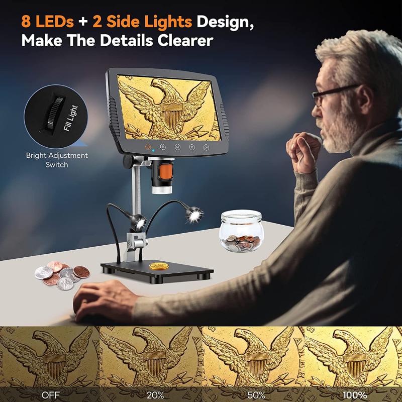
Light source and illumination are essential components of an optical microscope. The light source provides the necessary illumination to the specimen, allowing it to be viewed under the microscope. The illumination can be either transmitted or reflected, depending on the type of microscope and the specimen being observed.
In a transmitted light microscope, the light source is located beneath the stage and shines through the specimen. The light passes through the condenser, which focuses the light onto the specimen, and then through the objective lens, which magnifies the image. The image is then viewed through the eyepiece.
In a reflected light microscope, the light source is located above the stage and shines down onto the specimen. The light reflects off the specimen and is then collected by the objective lens and viewed through the eyepiece.
The type of illumination used can also affect the quality of the image. Brightfield illumination is the most common type of illumination and provides a bright, even background for the specimen. Darkfield illumination is used to observe specimens that are transparent or have low contrast, such as bacteria or small particles. Fluorescence illumination is used to observe specimens that emit light when exposed to specific wavelengths of light.
In recent years, advancements in technology have led to the development of new types of illumination, such as confocal microscopy and super-resolution microscopy. These techniques allow for even greater detail and resolution in the images produced by optical microscopes.
Overall, the light source and illumination are crucial components of an optical microscope, allowing for the observation and analysis of a wide range of specimens.
2、 Objective lens and magnification
Objective lens and magnification are two key components of an optical microscope that work together to produce a magnified image of a specimen. The objective lens is the lens closest to the specimen and is responsible for collecting and focusing the light that passes through the specimen. The magnification of the microscope is determined by the combination of the objective lens and the eyepiece lens.
When light passes through the objective lens, it is refracted and focused onto the specimen. The light then passes through the specimen and is refracted again as it passes through the objective lens. This produces an inverted and magnified image of the specimen.
The magnification of the microscope is determined by the focal length of the objective lens and the eyepiece lens. The total magnification is calculated by multiplying the magnification of the objective lens by the magnification of the eyepiece lens. For example, if the objective lens has a magnification of 10x and the eyepiece lens has a magnification of 20x, the total magnification of the microscope would be 200x.
In recent years, advancements in technology have led to the development of new types of objective lenses, such as super-resolution lenses, which can produce images with higher resolution and clarity. Additionally, digital microscopes have become increasingly popular, allowing for the capture and analysis of images using computer software.
Overall, the objective lens and magnification are essential components of an optical microscope, allowing for the visualization and analysis of specimens at a microscopic level.

3、 Stage and specimen preparation
Optical microscope how it works:
An optical microscope works by using visible light to magnify and resolve small objects. The light passes through the specimen and is then refracted by the lenses in the microscope to produce an enlarged image. The lenses in the microscope are designed to magnify the image and to correct for any distortions caused by the refractive properties of the specimen.
Stage and specimen preparation:
The stage of the microscope is where the specimen is placed for observation. The stage is typically a flat surface with clips or other mechanisms to hold the specimen in place. The specimen must be prepared in a way that allows it to be viewed under the microscope. This may involve staining the specimen to make it more visible or mounting it on a slide with a cover slip to protect it from damage.
In recent years, there has been a growing interest in using optical microscopy to study biological systems at the nanoscale. This has led to the development of new techniques for preparing specimens, such as cryo-electron microscopy, which allows specimens to be imaged at very low temperatures. Additionally, advances in computer technology have made it possible to process and analyze large amounts of data generated by optical microscopy, allowing researchers to gain new insights into the structure and function of biological systems.

4、 Eyepiece and image formation
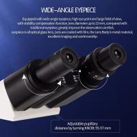
Eyepiece and image formation is a crucial aspect of how an optical microscope works. The eyepiece is the part of the microscope that the viewer looks through to observe the specimen. It contains a lens system that magnifies the image formed by the objective lens. The eyepiece typically has a magnification power of 10x, although this can vary depending on the microscope.
The image formation process begins with the objective lens, which is located near the specimen. The objective lens collects and focuses the light that passes through the specimen, creating a magnified image. This image is then projected through the body tube of the microscope and into the eyepiece.
The eyepiece contains a second lens system that further magnifies the image formed by the objective lens. This magnified image is what the viewer sees when looking through the eyepiece. The total magnification of the microscope is determined by multiplying the magnification of the objective lens by the magnification of the eyepiece.
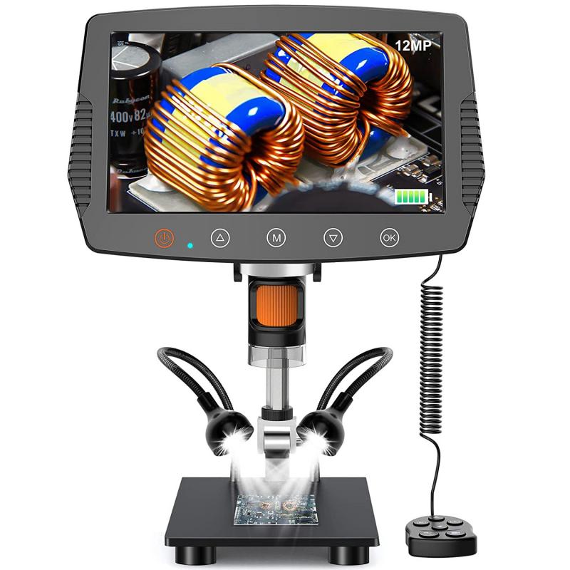
Recent advancements in technology have led to the development of digital microscopes, which use a camera to capture images of the specimen instead of relying on the viewer's eye. These microscopes can display the image on a computer screen, allowing for easier sharing and analysis of the specimen. Additionally, some digital microscopes use software to enhance the image, providing greater clarity and detail.
