What Are Different Types Of Microscope ?
There are several different types of microscopes, each with its own unique features and applications. Some common types include:
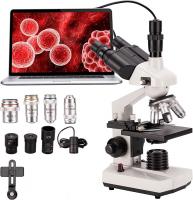
1. Optical Microscope: This is the most common type of microscope, which uses visible light to magnify and observe samples. It includes compound microscopes (with multiple lenses) and stereo microscopes (for 3D viewing).
2. Electron Microscope: These microscopes use a beam of electrons instead of light to create highly detailed images. There are two main types: transmission electron microscopes (TEM) and scanning electron microscopes (SEM).
3. Confocal Microscope: This type of microscope uses laser light to create high-resolution, 3D images of samples. It is commonly used in biological research and allows for optical sectioning of thick samples.
4. Scanning Probe Microscope: This microscope uses a physical probe to scan the surface of a sample, providing detailed information about its topography and properties. Examples include atomic force microscopes (AFM) and scanning tunneling microscopes (STM).
5. Fluorescence Microscope: These microscopes use fluorescent dyes to label specific structures or molecules within a sample, allowing for visualization of specific targets.
6. Phase Contrast Microscope: This type of microscope enhances the contrast of transparent samples by exploiting differences in refractive index.
These are just a few examples of the many types of microscopes available, each with its own advantages and applications in various scientific fields.
1、 Optical Microscope
The optical microscope is one of the most commonly used types of microscopes in scientific research and education. It uses visible light and a series of lenses to magnify and observe small objects or organisms. However, there are several different types of optical microscopes, each with its own unique features and applications.
1. Compound Microscope: This is the most basic and widely used type of optical microscope. It consists of two or more lenses that work together to magnify the specimen. Compound microscopes are commonly used in biology and medicine to observe cells, tissues, and small organisms.
2. Stereo Microscope: Also known as a dissecting microscope, this type of microscope provides a three-dimensional view of the specimen. It is commonly used in fields such as forensic science, entomology, and botany, where a larger working distance and depth perception are required.
3. Phase Contrast Microscope: This microscope is used to observe transparent or unstained specimens, such as living cells. It enhances the contrast between different parts of the specimen by exploiting the differences in refractive index.
4. Fluorescence Microscope: This microscope uses fluorescent dyes or proteins to label specific structures or molecules within the specimen. It allows researchers to visualize and study the distribution and behavior of these labeled components.
5. Confocal Microscope: This advanced microscope uses a laser beam to scan the specimen point by point, creating a three-dimensional image. It provides high-resolution images and is commonly used in biological research, particularly in studying cellular structures and processes.
6. Super-resolution Microscope: This recent development in microscopy allows for imaging beyond the diffraction limit of light. Techniques such as stimulated emission depletion (STED) microscopy and structured illumination microscopy (SIM) enable researchers to visualize structures at the nanoscale level.
In conclusion, the optical microscope encompasses various types, each with its own advantages and applications. From the basic compound microscope to the cutting-edge super-resolution microscope, these instruments have revolutionized our understanding of the microscopic world and continue to play a crucial role in scientific research and education.

2、 Electron Microscope
The Electron Microscope is one of the most powerful and advanced types of microscopes available today. It uses a beam of electrons instead of light to magnify and visualize objects at a much higher resolution than traditional light microscopes. This allows scientists to study the fine details of cells, tissues, and even individual atoms.
There are two main types of Electron Microscopes: Transmission Electron Microscope (TEM) and Scanning Electron Microscope (SEM). TEMs are used to study the internal structure of thin specimens, such as cells or tissues. They work by passing a beam of electrons through the specimen, which then interacts with the specimen and produces an image. This image can reveal the internal structure and composition of the specimen in great detail.
On the other hand, SEMs are used to study the surface of specimens. They work by scanning a focused beam of electrons across the specimen's surface and detecting the secondary electrons that are emitted. This creates a three-dimensional image of the specimen's surface, allowing scientists to study its topography and composition.
In recent years, there have been advancements in Electron Microscopy technology. For example, aberration-corrected Electron Microscopes have been developed, which minimize the distortions caused by the electron beam, resulting in even higher resolution images. Additionally, there have been improvements in the detection systems used in Electron Microscopes, allowing for faster and more sensitive imaging.
Overall, Electron Microscopes have revolutionized the field of microscopy, enabling scientists to explore the microscopic world with unprecedented detail and clarity. They continue to be an essential tool in various scientific disciplines, including biology, materials science, and nanotechnology.

3、 Confocal Microscope
A confocal microscope is one of the many types of microscopes used in scientific research and imaging. It is a powerful tool that allows scientists to obtain high-resolution images of biological samples with exceptional clarity and detail.
The confocal microscope works by using a laser beam to scan the sample point by point. It then collects the emitted light from each point and creates a three-dimensional image of the sample. This technique eliminates the out-of-focus light, resulting in sharper images with improved contrast and resolution compared to traditional microscopes.
In addition to its ability to produce high-resolution images, confocal microscopy also enables researchers to study live samples in real-time. This is achieved by using fluorescent dyes or proteins that can be specifically targeted to certain structures or molecules within the sample. By visualizing these fluorescent markers, scientists can track dynamic processes and observe cellular behavior in real-time.
Furthermore, confocal microscopy has been combined with other techniques to enhance its capabilities. For example, the integration of fluorescence lifetime imaging microscopy (FLIM) allows researchers to measure the fluorescence decay rate of molecules, providing valuable information about their environment and interactions.
In recent years, advancements in confocal microscopy have led to the development of super-resolution techniques such as stimulated emission depletion (STED) microscopy and structured illumination microscopy (SIM). These techniques surpass the diffraction limit of light, enabling researchers to visualize structures at the nanoscale level.
Overall, the confocal microscope is a versatile and powerful tool that continues to play a crucial role in various fields of research, including cell biology, neuroscience, and materials science. Its ability to provide high-resolution, real-time imaging has revolutionized our understanding of biological processes and has opened up new avenues for scientific discovery.

4、 Scanning Probe Microscope
A Scanning Probe Microscope (SPM) is a type of microscope that uses a physical probe to scan the surface of a sample to create an image with high resolution. It is capable of providing detailed information about the topography, electrical, magnetic, and mechanical properties of a sample at the nanoscale level.
There are several different types of SPMs, each with its own unique capabilities and applications. The most common types include:
1. Atomic Force Microscope (AFM): This type of SPM uses a small cantilever with a sharp tip to scan the surface of a sample. It measures the forces between the tip and the sample to create an image. AFM can provide information about surface roughness, mechanical properties, and even chemical composition.
2. Scanning Tunneling Microscope (STM): STM uses a conductive tip to scan the surface of a conductive sample. It measures the tunneling current between the tip and the sample to create an image. STM is particularly useful for studying the electronic properties of materials and can achieve atomic resolution.
3. Magnetic Force Microscope (MFM): MFM uses a magnetic tip to scan the surface of a sample. It measures the magnetic forces between the tip and the sample to create an image. MFM is commonly used to study magnetic materials and can provide information about their magnetic domains and properties.
4. Electrochemical Scanning Probe Microscope (EC-SPM): This type of SPM combines the capabilities of SPM with electrochemical measurements. It allows researchers to study the electrochemical properties of materials and investigate processes such as corrosion and electrochemical reactions.
In recent years, there have been advancements in SPM technology, such as the development of high-speed SPMs that can capture images at a faster rate, enabling real-time observations of dynamic processes. Additionally, there have been efforts to integrate SPM with other techniques, such as spectroscopy, to obtain more comprehensive information about the sample.
Overall, SPMs have revolutionized the field of nanoscience and nanotechnology by providing researchers with the ability to visualize and manipulate matter at the atomic and molecular level. They continue to play a crucial role in various scientific disciplines, including materials science, physics, chemistry, and biology.


