What Are The Main Types Of Microscope ?
The main types of microscopes include optical microscopes, electron microscopes, and scanning probe microscopes.
1、 Optical Microscope
The main types of microscopes are optical microscopes, electron microscopes, and scanning probe microscopes. Optical microscopes, also known as light microscopes, are the most commonly used type of microscope. They use visible light and a series of lenses to magnify and observe small objects or organisms. Optical microscopes can be further classified into several subtypes, including compound microscopes, stereo microscopes, and digital microscopes.
Compound microscopes are the most traditional type of optical microscope and are used to view thin, transparent specimens. They have two sets of lenses, an objective lens near the specimen and an eyepiece lens near the observer's eye. Stereo microscopes, on the other hand, provide a three-dimensional view of larger, opaque specimens. They have two separate optical paths for each eye, allowing for depth perception.
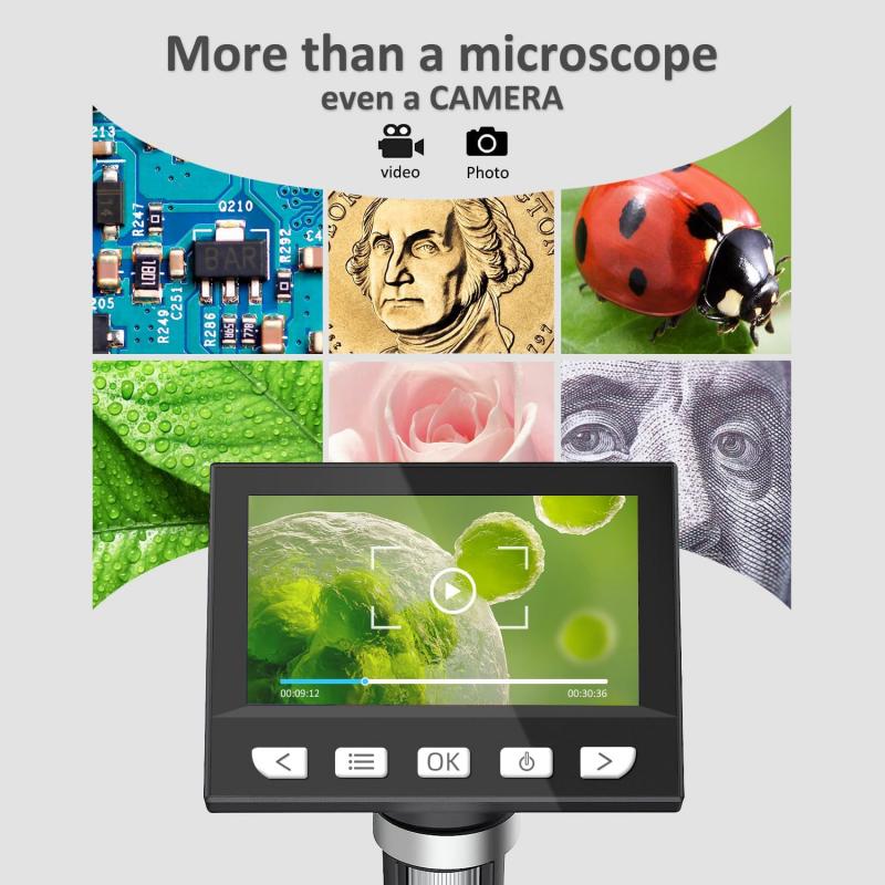
Digital microscopes are a more recent development in optical microscopy. They use a digital camera to capture images of the specimen, which can then be viewed on a computer screen or other digital display. This allows for easy sharing and analysis of the images.
In recent years, there have been advancements in optical microscopy techniques, such as confocal microscopy and super-resolution microscopy. Confocal microscopy uses a laser to scan the specimen point by point, creating a series of optical sections that can be reconstructed into a three-dimensional image. Super-resolution microscopy techniques, such as stimulated emission depletion (STED) microscopy and structured illumination microscopy (SIM), allow for imaging beyond the diffraction limit of light, providing higher resolution and more detailed images.
Overall, optical microscopes continue to be an essential tool in various scientific fields, including biology, medicine, materials science, and more. The advancements in technology have allowed for improved imaging capabilities and have expanded the range of applications for optical microscopy.

2、 Electron Microscope
The main types of microscopes are light microscopes and electron microscopes. Light microscopes use visible light to illuminate the specimen and magnify it, allowing for the observation of cells, tissues, and small organisms. They are commonly used in biology and medicine for routine laboratory work and research.
On the other hand, electron microscopes use a beam of electrons instead of light to magnify the specimen. There are two main types of electron microscopes: transmission electron microscopes (TEM) and scanning electron microscopes (SEM). TEMs use a thin section of the specimen and transmit electrons through it to create a detailed image of the internal structure. SEMs, on the other hand, scan the surface of the specimen with a focused beam of electrons to create a three-dimensional image.
Electron microscopes have revolutionized the field of microscopy by providing much higher resolution and magnification capabilities compared to light microscopes. They can reveal intricate details of cells, molecules, and even individual atoms. This has led to significant advancements in various scientific disciplines, including materials science, nanotechnology, and biology.
In recent years, there have been advancements in electron microscopy techniques, such as cryo-electron microscopy (cryo-EM). Cryo-EM allows for the imaging of biological samples in their native, frozen state, providing high-resolution structural information of biomolecules. This technique has been instrumental in understanding the structure and function of complex biological systems, including proteins and viruses.
Overall, electron microscopes, including TEMs and SEMs, have become indispensable tools in scientific research, enabling scientists to explore the microscopic world with unprecedented detail and clarity.

3、 Confocal Microscope
The main types of microscopes include the optical microscope, electron microscope, and the confocal microscope. The confocal microscope is a powerful tool used in various scientific fields, including biology, medicine, and materials science.
A confocal microscope uses a laser beam to scan a sample point by point, creating a three-dimensional image. It offers several advantages over traditional microscopes, such as improved resolution, optical sectioning, and the ability to capture images at different depths within a sample. This allows researchers to study the structure and function of cells and tissues in great detail.
In recent years, confocal microscopy has seen advancements in technology, leading to improved imaging capabilities. For example, the development of super-resolution techniques, such as stimulated emission depletion (STED) microscopy and structured illumination microscopy (SIM), has allowed researchers to achieve resolutions beyond the diffraction limit of light. This has opened up new possibilities for studying subcellular structures and processes with unprecedented detail.
Furthermore, confocal microscopy has been combined with other imaging techniques, such as fluorescence lifetime imaging microscopy (FLIM) and fluorescence resonance energy transfer (FRET), to provide additional information about molecular interactions and dynamics within cells. These advancements have greatly expanded the applications of confocal microscopy in fields like neuroscience, cancer research, and drug discovery.
In conclusion, the confocal microscope is one of the main types of microscopes used in scientific research. Its ability to provide high-resolution, three-dimensional images has revolutionized the study of cells and tissues. With ongoing technological advancements, confocal microscopy continues to evolve, enabling researchers to delve deeper into the intricate world of biology and medicine.

4、 Scanning Probe Microscope
The main types of microscopes include optical microscopes, electron microscopes, and scanning probe microscopes. Optical microscopes use visible light to magnify and observe samples, while electron microscopes use a beam of electrons to achieve higher magnification and resolution. Scanning probe microscopes (SPMs) are a more recent addition to the field of microscopy and offer unique capabilities.
SPMs are a family of microscopes that use a physical probe to scan the surface of a sample and create an image with atomic-scale resolution. The most common types of SPMs are atomic force microscopes (AFMs) and scanning tunneling microscopes (STMs). AFMs use a small probe with a sharp tip to measure the forces between the tip and the sample surface, providing information about the topography and mechanical properties of the sample. STMs, on the other hand, rely on the quantum tunneling effect to measure the current flowing between the tip and the sample, providing information about the electronic properties of the sample.
One of the latest developments in SPM technology is the combination of AFM and STM capabilities into a single instrument called a scanning probe microscope. This hybrid microscope allows researchers to simultaneously obtain topographic and electronic information about a sample, providing a more comprehensive understanding of its properties. Additionally, advancements in SPM technology have led to the development of new imaging modes, such as Kelvin probe force microscopy and magnetic force microscopy, which enable the investigation of electrical and magnetic properties at the nanoscale.
Overall, scanning probe microscopes have revolutionized the field of microscopy by enabling researchers to study and manipulate materials at the atomic and molecular level. Their unique capabilities make them invaluable tools in various scientific disciplines, including materials science, nanotechnology, and biology.


