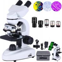
What Can I See With 1000x Microscope ?
With a 1000x microscope, you can see a wide range of microscopic objects and details. You can observe cells, bacteria, fungi, and other microorganisms in great detail. Additionally, you can examine the structure of plant and animal tissues, such as the arrangement of cells in leaves or the organization of muscle fibers. You may also be able to visualize the intricate patterns and textures of minerals and crystals. Furthermore, you can explore the world of microorganisms in pond water, discovering various organisms like amoebas, paramecia, and rotifers. Overall, a 1000x microscope allows you to delve into the fascinating realm of the microscopic world and explore the intricate details of various specimens.
1、 Cellular Structures and Organelles
With a 1000x microscope, you can observe a wide range of cellular structures and organelles in great detail. This level of magnification allows for the visualization of intricate features that are not visible to the naked eye. Here are some of the cellular structures and organelles that you can see:
1. Nucleus: The control center of the cell, containing genetic material (DNA) and surrounded by a nuclear membrane. You can observe the nucleolus, chromatin, and nuclear pores within the nucleus.
2. Mitochondria: These organelles are responsible for energy production in the cell. With a 1000x microscope, you can see the inner and outer mitochondrial membranes, cristae (folds), and the matrix.
3. Endoplasmic reticulum (ER): This network of membranes is involved in protein synthesis and lipid metabolism. You can observe rough ER (studded with ribosomes) and smooth ER (lacking ribosomes).
4. Golgi apparatus: This organelle modifies, sorts, and packages proteins for transport. With a 1000x microscope, you can see the stacked cisternae and vesicles.
5. Lysosomes: These organelles contain digestive enzymes and are involved in breaking down waste materials. You can observe the membrane-bound vesicles and the dense contents within.
6. Cytoskeleton: This network of protein filaments provides structural support and facilitates cell movement. With a 1000x microscope, you can see microtubules, microfilaments, and intermediate filaments.
7. Plasma membrane: The outer boundary of the cell, composed of a phospholipid bilayer. You can observe the lipid bilayer and embedded proteins.
It is important to note that the latest advancements in microscopy techniques, such as super-resolution microscopy, have pushed the limits of resolution beyond the traditional optical microscope. These techniques allow for the visualization of even smaller cellular structures and molecular interactions.

2、 Microorganisms and Bacteria
With a 1000x microscope, you can observe a wide range of microorganisms and bacteria that are otherwise invisible to the naked eye. This level of magnification allows for detailed examination of their structures, behaviors, and interactions.
One of the most common microorganisms that can be observed with a 1000x microscope is bacteria. Bacteria are single-celled organisms that exist in various shapes and sizes. They can be found virtually everywhere, from soil and water to the human body. With this microscope, you can study the different types of bacteria, such as cocci (spherical), bacilli (rod-shaped), and spirilla (spiral-shaped). You can also observe their cellular structures, including the cell wall, cytoplasm, and genetic material.
Apart from bacteria, a 1000x microscope allows you to explore other microorganisms like protozoa, algae, and fungi. Protozoa are single-celled eukaryotic organisms that exhibit diverse shapes and locomotion methods. Algae, on the other hand, are photosynthetic microorganisms that can be found in aquatic environments. With this microscope, you can observe their chloroplasts and cell structures.
Furthermore, a 1000x microscope enables you to study the intricate details of viruses. Viruses are smaller than bacteria and can only be observed using an electron microscope. However, some larger viruses can be visualized with a 1000x microscope. By examining viruses, scientists can gain insights into their structure, replication mechanisms, and potential treatments.
It is important to note that the field of microbiology is constantly evolving, and new discoveries are being made regularly. With advancements in microscopy techniques and technology, scientists are able to observe microorganisms and bacteria in greater detail than ever before. This allows for a deeper understanding of their biology, ecology, and impact on human health.

3、 Tissue and Cell Morphology
With a 1000x microscope, you can observe and study various aspects of tissue and cell morphology in great detail. This level of magnification allows for a closer examination of cellular structures and their functions.
At this magnification, you can observe the intricate details of cell organelles such as the nucleus, mitochondria, endoplasmic reticulum, Golgi apparatus, and lysosomes. You can also observe the cytoskeleton, which includes microtubules, microfilaments, and intermediate filaments, providing structural support to the cell. Additionally, you can study the cell membrane and its components, including proteins and lipids.
Furthermore, a 1000x microscope enables the observation of tissue morphology. You can examine the arrangement and organization of cells within tissues, such as epithelial, connective, muscle, and nervous tissues. This level of magnification allows for the identification of different cell types within a tissue and the observation of their specific functions.
In recent years, advancements in microscopy techniques have allowed for even more detailed observations. For example, the use of fluorescent dyes and markers can help visualize specific cellular components or processes. Additionally, techniques such as confocal microscopy and super-resolution microscopy have further enhanced the resolution and clarity of cellular and tissue imaging.
Overall, a 1000x microscope provides a powerful tool for studying tissue and cell morphology. It allows researchers to explore the intricate structures and functions of cells, contributing to our understanding of various biological processes and diseases.

4、 Blood Cells and Circulatory System
With a 1000x microscope, you can observe various components of the blood cells and the circulatory system in great detail. The high magnification allows for a closer examination of the intricate structures and functions of these vital elements of the human body.
When observing blood cells, you can see the different types of cells present in the bloodstream. Red blood cells, also known as erythrocytes, appear as small, biconcave discs. Their main function is to transport oxygen from the lungs to the body's tissues. With the microscope, you can observe their unique shape and the absence of a nucleus, which allows for more space to carry oxygen.
White blood cells, or leukocytes, are another important component of the circulatory system. They play a crucial role in the immune response, defending the body against infections and foreign substances. With the microscope, you can observe the various types of white blood cells, such as neutrophils, lymphocytes, monocytes, eosinophils, and basophils. Each type has distinct characteristics and functions, which can be observed and studied at high magnification.
Additionally, platelets, also known as thrombocytes, can be observed with a 1000x microscope. These small, irregularly shaped cells are responsible for blood clotting, preventing excessive bleeding when a blood vessel is damaged. You can observe their role in forming clumps and initiating the clotting process.
Furthermore, the microscope can provide insights into the structure and function of blood vessels. Capillaries, the smallest blood vessels, can be observed in detail, revealing their thin walls and the exchange of oxygen, nutrients, and waste products between the blood and surrounding tissues.
In recent years, advancements in microscopy techniques have allowed for even more detailed observations. For example, fluorescence microscopy can be used to label specific molecules or structures within the blood cells, providing a clearer understanding of their functions and interactions. Additionally, confocal microscopy enables the visualization of three-dimensional structures, allowing for a more comprehensive analysis of the circulatory system.
In conclusion, with a 1000x microscope, you can observe and study the intricate details of blood cells and the circulatory system. This level of magnification provides valuable insights into their structure, function, and interactions, contributing to our understanding of human physiology and the diagnosis and treatment of various diseases.


There are no comments for this blog.