What Is A Microscope Simple Definition ?
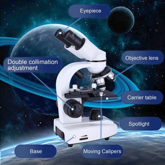
A microscope is a scientific instrument used to magnify and observe objects that are too small to be seen with the naked eye. It consists of a combination of lenses and sometimes other optical components that work together to produce a magnified image of the specimen being examined. Microscopes are widely used in various fields of science, such as biology, medicine, and materials science, to study and analyze microscopic structures and organisms.
1、 Optical Microscopy: Using lenses to magnify and observe small objects.
A microscope is a scientific instrument that is used to magnify and observe small objects that are not visible to the naked eye. It works by using lenses to enhance the resolution and magnification of the object being observed. The most common type of microscope is the optical microscope, which utilizes visible light to illuminate the specimen.
Optical microscopy involves the use of lenses to focus light onto the specimen, allowing for a detailed examination of its structure and composition. The light passes through the specimen and is then magnified by a series of lenses, ultimately forming an enlarged image that can be observed by the human eye or captured using a camera.
In recent years, there have been advancements in optical microscopy techniques, such as confocal microscopy and fluorescence microscopy. Confocal microscopy uses a laser to scan the specimen in a series of thin optical sections, resulting in a three-dimensional image with improved resolution. Fluorescence microscopy involves the use of fluorescent dyes or proteins that emit light when excited by specific wavelengths, allowing for the visualization of specific structures or molecules within the specimen.
Furthermore, there have been developments in digital microscopy, where the images obtained from the microscope are directly captured by a digital camera and displayed on a computer screen. This enables the storage, analysis, and sharing of microscopic images in a more convenient and efficient manner.
Overall, optical microscopy remains a fundamental tool in various scientific disciplines, including biology, medicine, materials science, and forensics, providing valuable insights into the microscopic world.

2、 Electron Microscopy: Using electron beams to visualize tiny structures.
A microscope is a scientific instrument that is used to magnify and visualize objects that are too small to be seen with the naked eye. It works by using a combination of lenses and light to enlarge the image of the object being observed. Microscopes are commonly used in various fields of science, such as biology, medicine, and materials science, to study and analyze microscopic structures and organisms.
Electron microscopy is a specialized type of microscopy that utilizes electron beams instead of light to visualize tiny structures. It offers much higher magnification and resolution compared to traditional light microscopy. Instead of using lenses, electron microscopes use electromagnetic lenses to focus the electron beam onto the specimen. The interaction between the electrons and the specimen produces signals that are then detected and used to create an image.
Electron microscopy has revolutionized our understanding of the microscopic world. It has allowed scientists to observe and study structures at the atomic and molecular level, providing valuable insights into the composition, structure, and behavior of materials and biological specimens. This technique has been instrumental in advancing fields such as nanotechnology, materials science, and cell biology.
In recent years, advancements in electron microscopy have further improved its capabilities. Techniques such as scanning electron microscopy (SEM) and transmission electron microscopy (TEM) have become more sophisticated, allowing for even higher resolution imaging and the ability to study dynamic processes in real-time. Additionally, the development of cryo-electron microscopy (cryo-EM) has enabled the imaging of biological samples in their native, hydrated state, providing unprecedented details of cellular structures and molecular interactions.
Overall, electron microscopy continues to be a powerful tool in scientific research, enabling scientists to explore the intricate world of the small and uncover new knowledge about the building blocks of life and matter.

3、 Compound Microscope: A microscope with multiple lenses for higher magnification.
A compound microscope is a scientific instrument that uses multiple lenses to magnify small objects or specimens for observation. It is one of the most commonly used types of microscopes in various scientific fields, including biology, medicine, and materials science.
The basic design of a compound microscope consists of two optical systems: the objective lens and the eyepiece. The objective lens is located near the specimen and is responsible for gathering light and forming a magnified image. The eyepiece, or ocular lens, is positioned at the top of the microscope and further magnifies the image formed by the objective lens, allowing the viewer to see a highly magnified and detailed view of the specimen.
Compound microscopes typically have multiple objective lenses with different magnification powers, allowing users to switch between different levels of magnification depending on their needs. The combination of these lenses enables the microscope to achieve higher magnification than a simple microscope, which only has one lens.
In recent years, advancements in technology have led to the development of more sophisticated compound microscopes with features such as digital imaging capabilities, allowing users to capture and analyze images of specimens on a computer screen. Additionally, some compound microscopes now incorporate advanced techniques like fluorescence microscopy, confocal microscopy, and phase contrast microscopy, which enhance the visualization of specific structures or processes within the specimen.
Overall, compound microscopes play a crucial role in scientific research, education, and various industries by enabling scientists and professionals to study and understand the intricate details of microscopic objects and phenomena.

4、 Scanning Probe Microscopy: Imaging surfaces using a physical probe.
Scanning Probe Microscopy (SPM) is a powerful technique used to image and study surfaces at the nanoscale level. It involves the use of a physical probe to scan and interact with the surface of a sample, providing detailed information about its topography, properties, and composition.
The basic principle of SPM is to bring a sharp probe very close to the surface of the sample and measure the interaction between the probe and the surface. The probe is typically a sharp tip made of a conductive material, such as silicon or metal, which is mounted on a cantilever. As the probe scans the surface, it moves up and down in response to the forces between the tip and the sample. These movements are detected and used to generate an image of the surface.
SPM techniques include Atomic Force Microscopy (AFM), Scanning Tunneling Microscopy (STM), and many others. AFM is widely used and can provide high-resolution images of various materials, including biological samples. STM, on the other hand, is particularly useful for studying conductive materials and can even reveal individual atoms on a surface.
The latest advancements in SPM have allowed for not only imaging but also manipulation of individual atoms and molecules. This has opened up new possibilities in fields such as nanotechnology, materials science, and biology. SPM has become an indispensable tool for researchers and scientists seeking to understand and manipulate matter at the nanoscale level.
In summary, Scanning Probe Microscopy is a technique that uses a physical probe to scan and interact with the surface of a sample, providing detailed information about its topography and properties. It has revolutionized our ability to study and manipulate matter at the nanoscale, leading to numerous advancements in various scientific disciplines.


