What Is A Specimen Microscope ?
A specimen microscope is a type of microscope that is designed to observe small specimens, such as cells or microorganisms, at high magnification. It typically consists of a light source, a stage for holding the specimen, and a series of lenses that magnify the image of the specimen. The microscope may also have additional features, such as a camera or digital imaging system, to capture images of the specimen for further analysis or documentation. Specimen microscopes are commonly used in scientific research, medical diagnosis, and education, and are available in a range of sizes and configurations to suit different applications.
1、 Definition and Function of Specimen Microscope

A specimen microscope is a type of microscope that is designed to observe and analyze small specimens such as cells, tissues, and microorganisms. It is also known as a biological microscope or a compound microscope. The specimen microscope uses a combination of lenses to magnify the specimen and provide a clear image for observation.
The function of a specimen microscope is to allow scientists and researchers to study the structure and function of living organisms at a microscopic level. This type of microscope is commonly used in biology, medicine, and other life sciences to study the structure and function of cells, tissues, and microorganisms.
The specimen microscope has evolved over time, with advancements in technology leading to improvements in magnification, resolution, and imaging capabilities. Modern specimen microscopes often include digital imaging technology, allowing researchers to capture and analyze images of specimens in real-time.
In addition to its use in scientific research, the specimen microscope is also used in medical diagnosis and treatment. Pathologists use specimen microscopes to examine tissue samples for signs of disease, while surgeons use them to guide delicate procedures.
Overall, the specimen microscope is an essential tool in the study of life sciences, providing researchers with a powerful tool for observing and analyzing the microscopic world.
2、 Types of Specimen Microscopes (e.g. Compound, Stereo, Digital)

Types of Specimen Microscopes (e.g. Compound, Stereo, Digital)
A specimen microscope is a type of microscope that is used to observe small specimens such as cells, tissues, and microorganisms. There are several types of specimen microscopes available, each with its own unique features and capabilities.
Compound microscopes are the most common type of specimen microscope. They use a series of lenses to magnify the specimen, and are capable of magnifying up to 1000 times. Compound microscopes are commonly used in biology and medical research.
Stereo microscopes, also known as dissecting microscopes, are used to observe larger specimens such as insects, rocks, and plants. They provide a three-dimensional view of the specimen, making it easier to observe its structure and features.
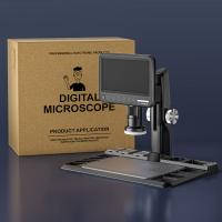
Digital microscopes are a newer type of specimen microscope that use digital imaging technology to capture images of the specimen. They are often used in research and education, as they allow for easy sharing and analysis of images.
In recent years, there has been a growing interest in portable and handheld specimen microscopes. These microscopes are small and lightweight, making them ideal for fieldwork and on-the-go observations. They are often used in environmental science, geology, and archaeology.
Overall, the type of specimen microscope used will depend on the specific needs of the researcher or observer. Advances in technology have led to the development of new and innovative types of specimen microscopes, making it easier than ever to observe and study the microscopic world.
3、 Components of a Specimen Microscope (e.g. Eyepiece, Objective Lens, Stage)

A specimen microscope is a type of microscope that is used to observe small specimens such as cells, tissues, and microorganisms. It is designed to provide high magnification and resolution, allowing scientists and researchers to study the structure and function of these specimens in detail.
The components of a specimen microscope include the eyepiece, objective lens, stage, focus knobs, and light source. The eyepiece is the part of the microscope that the user looks through to observe the specimen. The objective lens is the lens closest to the specimen and provides the primary magnification. The stage is the platform on which the specimen is placed for observation. The focus knobs are used to adjust the focus of the microscope, while the light source provides illumination for the specimen.
In recent years, there have been advancements in specimen microscope technology, including the development of digital microscopes. These microscopes use digital cameras to capture images of the specimen, which can then be viewed on a computer screen or other digital device. This technology has made it easier for researchers to share and analyze microscope images, as well as to store and organize large amounts of data.
Overall, the specimen microscope remains an essential tool for scientific research and discovery, allowing scientists to explore the microscopic world in detail and gain a deeper understanding of the natural world.
4、 Techniques for Preparing Specimens for Microscopy (e.g. Staining, Mounting)

What is a specimen microscope?
A specimen microscope is a type of microscope that is specifically designed for the observation of specimens. It is used to magnify and examine small objects, such as cells, tissues, and microorganisms. Specimen microscopes are commonly used in biology, medicine, and other scientific fields.
Techniques for Preparing Specimens for Microscopy (e.g. Staining, Mounting)
There are several techniques for preparing specimens for microscopy, including staining and mounting. Staining is a process that involves adding a dye or stain to a specimen to enhance its visibility under a microscope. Different stains are used for different types of specimens, and the choice of stain depends on the type of specimen being examined.
Mounting is the process of placing a specimen on a slide and covering it with a cover slip. This helps to protect the specimen and keep it in place while it is being examined under the microscope. Mounting can be done using a variety of different techniques, including wet mounts, dry mounts, and permanent mounts.
In recent years, there has been a growing interest in new techniques for preparing specimens for microscopy, such as 3D imaging and super-resolution microscopy. These techniques allow for more detailed and accurate imaging of specimens, and are helping to advance our understanding of the microscopic world.
















There are no comments for this blog.