What Is Low Magnification On A Microscope ?
Low magnification on a microscope refers to the setting or objective lens that provides a lower level of magnification. It allows for a wider field of view, enabling the observation of larger specimens or structures. This setting is typically used when a broader overview or initial examination of a sample is required before moving on to higher magnifications for more detailed analysis.
1、 Definition and Purpose of Low Magnification in Microscopy
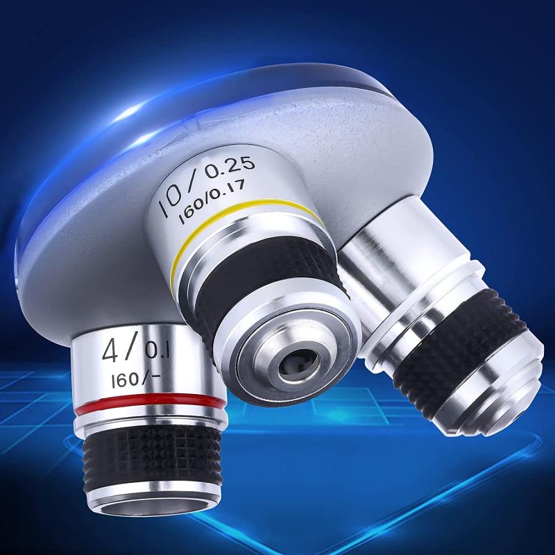
Low magnification on a microscope refers to the setting or objective lens that provides a lower level of magnification compared to higher magnification settings. It typically ranges from 4x to 10x, although the exact magnification may vary depending on the microscope model.
The purpose of using low magnification in microscopy is to obtain a wider field of view and a larger depth of field. This allows for a broader perspective and a better understanding of the overall structure and organization of the specimen being observed. Low magnification is particularly useful when studying larger specimens or when trying to locate specific areas of interest within a sample.
By using low magnification, researchers can quickly scan a sample and identify regions that require further examination at higher magnifications. It helps in locating specific structures or features of interest, such as cells, tissues, or organisms, and provides a context for subsequent higher magnification observations.
In recent years, there has been an increasing emphasis on the use of low magnification in microscopy for various applications. For example, in the field of pathology, low magnification is used to assess the overall architecture of tissues and identify abnormal areas that may require further investigation. In materials science, low magnification is employed to study the macroscopic properties and structures of materials.
Overall, low magnification on a microscope serves as a valuable tool for obtaining a broader understanding of a specimen's organization and structure. It aids in guiding subsequent higher magnification observations and provides a more comprehensive analysis of the sample under study.

2、 Advantages and Limitations of Low Magnification in Microscopy
Low magnification on a microscope refers to the use of lower levels of magnification to observe specimens. It typically ranges from 10x to 40x, allowing for a wider field of view and a larger area of the specimen to be seen at once. This level of magnification is commonly used in routine laboratory work, educational settings, and initial observations of samples.
One advantage of low magnification is that it provides a broader perspective of the specimen. It allows for the observation of the overall structure, shape, and arrangement of cells or organisms. This is particularly useful when studying larger specimens or when trying to identify general characteristics of a sample. Additionally, low magnification enables faster scanning of a larger area, which can be beneficial when searching for specific features or abnormalities.
Another advantage is that low magnification often requires less time and effort to prepare samples. Specimens can be observed directly without the need for complex staining or sectioning techniques. This makes it a cost-effective and time-efficient method for routine examinations.
However, low magnification also has its limitations. One major limitation is the reduced level of detail that can be observed. Fine structures and smaller components may not be visible at this level of magnification, making it unsuitable for detailed analysis or identification of specific cellular or subcellular features. In such cases, higher magnification levels are required.
Moreover, low magnification may not be suitable for studying certain types of specimens, such as transparent or translucent samples. These samples may require higher magnification and specialized techniques, such as phase contrast or differential interference contrast microscopy, to enhance visibility.
In recent years, there has been a growing interest in the use of low magnification microscopy for whole-slide imaging and digital pathology. This approach involves scanning entire slides at low magnification and then digitally zooming in on specific regions of interest for detailed examination. This technique allows for efficient storage, sharing, and analysis of large amounts of data, making it particularly useful in research and diagnostic settings.
In conclusion, low magnification on a microscope provides a broader perspective and faster scanning of specimens, making it suitable for routine examinations and initial observations. However, it has limitations in terms of detail and suitability for certain types of samples. The latest point of view highlights the emerging use of low magnification microscopy in whole-slide imaging and digital pathology, which offers new possibilities for efficient data analysis and storage.
3、 Applications of Low Magnification in Microscopy
Low magnification on a microscope refers to the setting or objective lens that provides a lower level of magnification compared to higher magnification settings. It allows for a wider field of view, enabling the observation of larger specimens or a broader area of a sample. Low magnification is typically used when a general overview or initial examination of a sample is required before moving on to higher magnification settings.
Applications of low magnification in microscopy are diverse and play a crucial role in various scientific fields. In biology, low magnification is often used to study the overall structure and organization of tissues or organs. It allows researchers to observe the arrangement of cells, identify different cell types, and understand the overall architecture of a biological sample. This is particularly useful in histology, where low magnification helps in identifying abnormal tissue structures or lesions.
In materials science, low magnification microscopy is employed to examine the surface morphology and texture of materials. It aids in identifying defects, cracks, or impurities in a sample, which can provide valuable insights into the material's properties and quality. Additionally, low magnification is used in quality control processes to assess the uniformity and consistency of manufactured products.
In recent years, there has been an increasing interest in using low magnification microscopy for environmental monitoring and analysis. It allows for the examination of larger environmental samples, such as soil or water, to study the distribution of microorganisms, pollutants, or other particles. This approach provides a broader perspective on the ecological health of an environment and aids in identifying potential threats or changes in biodiversity.
Furthermore, low magnification microscopy is also utilized in educational settings to introduce students to the basics of microscopy and to develop their observational skills. It allows beginners to grasp the concept of microscopy and understand the importance of different magnification levels in scientific research.
In conclusion, low magnification on a microscope is essential for obtaining a wider field of view and is used in various scientific fields. Its applications range from studying biological tissues and materials to environmental monitoring and educational purposes. As technology advances, there may be further developments in low magnification microscopy techniques, enabling more precise and detailed observations at lower magnification levels.
4、 Techniques and Methods for Achieving Low Magnification in Microscopy
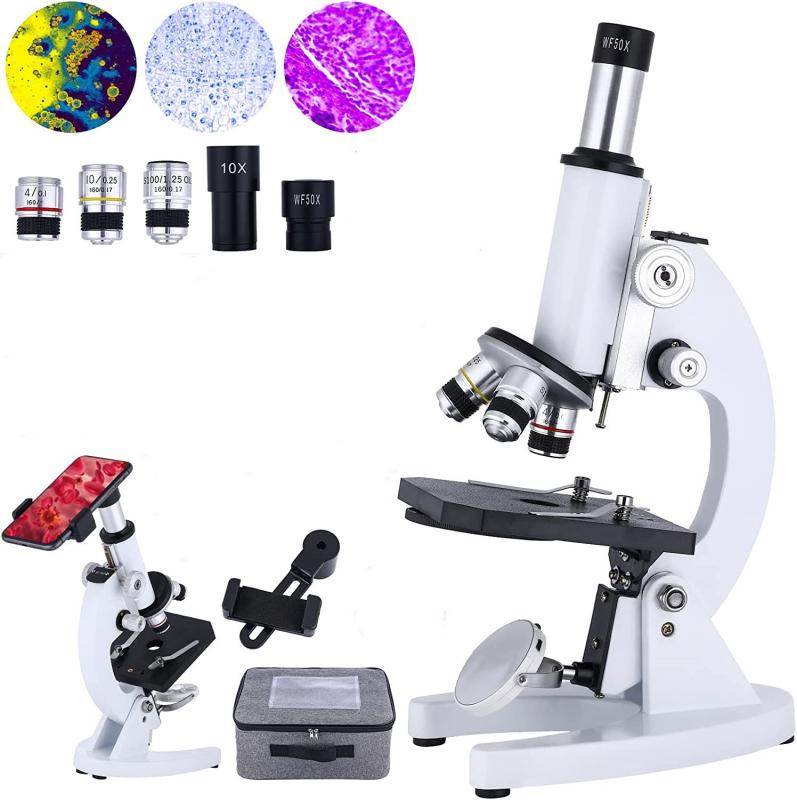
Low magnification on a microscope refers to the use of lower power objectives or lenses to observe specimens at a lower level of detail. It allows for a wider field of view, enabling researchers to observe larger areas of a sample or specimen. This technique is particularly useful when studying macroscopic features or when trying to get an overall view of a sample before zooming in for more detailed analysis.
To achieve low magnification, microscopes typically have objectives with lower numerical aperture (NA) and longer focal lengths. These objectives have a lower magnification power, usually ranging from 2x to 10x, compared to higher magnification objectives that can go up to 100x or more. Additionally, low magnification objectives often have a larger working distance, which is the distance between the objective lens and the specimen. This allows for the observation of thicker samples or specimens that require more space.
In recent years, there has been an increasing interest in low magnification microscopy due to its potential applications in various fields. For example, in materials science, low magnification microscopy can be used to study the overall structure and composition of materials, such as metals or polymers. In biology, it can be employed to observe larger organisms or tissues, such as plant leaves or animal organs.
Furthermore, advancements in imaging technology have allowed for the development of new techniques to achieve low magnification. For instance, wide-field microscopy techniques, such as brightfield or phase contrast microscopy, can provide a larger field of view without the need for high magnification objectives. Additionally, techniques like panoramic imaging or stitching can be used to create high-resolution images of large areas by combining multiple low magnification images.
In conclusion, low magnification on a microscope refers to the use of lower power objectives to observe specimens at a wider field of view. This technique is valuable for studying macroscopic features or obtaining an overall view of a sample. With the advancements in imaging technology, low magnification microscopy has gained importance in various fields, offering new possibilities for research and analysis.












There are no comments for this blog.