What Power Microscope To See Blood Cells ?
A compound light microscope with a magnification of at least 400x is required to see blood cells. This type of microscope uses visible light and a series of lenses to magnify the sample. Blood cells are very small, with red blood cells typically measuring around 7-8 micrometers in diameter and white blood cells ranging from 10-20 micrometers. Therefore, a high magnification is necessary to observe them in detail. Additionally, a microscope with a good resolution is important to distinguish the different types of blood cells and their structures. It is also recommended to use a microscope with a built-in light source or an external light source to illuminate the sample and enhance the contrast.
1、 Compound Microscopes
The best power microscope to see blood cells is a compound microscope. Compound microscopes are the most commonly used type of microscope in biology and medical laboratories. They use two or more lenses to magnify the image of the specimen, allowing for a clear and detailed view of the blood cells.
Compound microscopes are capable of magnifying the image of the specimen up to 1000 times, which is sufficient to see the blood cells. They are also equipped with high-quality lenses that provide a clear and sharp image of the specimen. Additionally, compound microscopes are easy to use and require minimal training, making them ideal for use in medical and biology laboratories.
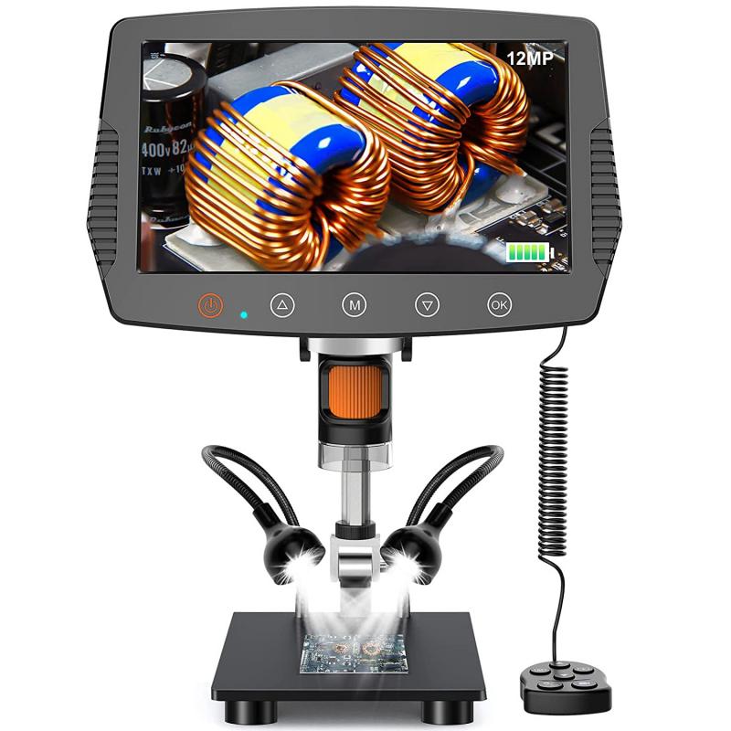
In recent years, there has been a growing interest in using digital microscopes to view blood cells. Digital microscopes use a camera to capture images of the specimen, which can then be viewed on a computer screen. This technology has several advantages over traditional compound microscopes, including the ability to share images with others and the ability to store images for future reference.
However, digital microscopes are still relatively new and may not be as widely available as traditional compound microscopes. Additionally, they may be more expensive than traditional microscopes, which could limit their use in some settings.
Overall, the best power microscope to see blood cells is a compound microscope. While digital microscopes offer some advantages, traditional compound microscopes remain the most widely used and accessible option for viewing blood cells in medical and biology laboratories.

2、 Brightfield Microscopy
The power microscope required to see blood cells is a Brightfield Microscope. This type of microscope is commonly used in medical laboratories and research facilities to observe and analyze biological specimens, including blood cells. Brightfield microscopy is a simple and widely used technique that involves illuminating the sample with a bright light source and observing the specimen through the objective lens.
In a Brightfield Microscope, the light passes through the specimen and is absorbed or scattered by the cells, creating contrast between the cells and the surrounding medium. This allows for the visualization of the cells and their structures, such as the nucleus, cytoplasm, and cell membrane. The magnification of the microscope can be adjusted to provide a clear and detailed view of the cells.
Recent advancements in microscopy technology have led to the development of more advanced techniques, such as fluorescence microscopy and confocal microscopy. These techniques offer higher resolution and greater specificity for certain types of samples, but they are not always necessary for observing blood cells. Brightfield microscopy remains a reliable and effective method for observing blood cells and is still widely used in medical and research settings.
In conclusion, a Brightfield Microscope is the power microscope required to see blood cells. This technique is simple, reliable, and widely used in medical and research laboratories. While newer microscopy techniques have been developed, Brightfield microscopy remains an essential tool for observing blood cells and other biological specimens.

3、 Phase Contrast Microscopy
The power microscope required to see blood cells is a Phase Contrast Microscope. This type of microscope is specifically designed to enhance the contrast of transparent and colorless specimens, such as blood cells, by converting the differences in refractive index into variations in light intensity. This allows for the visualization of the cells without the need for staining or other sample preparation techniques.
Phase Contrast Microscopy has been widely used in the field of hematology for the examination of blood cells. It allows for the identification and analysis of different types of blood cells, including red blood cells, white blood cells, and platelets. This technique has also been used to study the morphology and behavior of blood cells in various diseases, such as leukemia and sickle cell anemia.
In recent years, there has been a growing interest in the use of Phase Contrast Microscopy for the diagnosis of malaria. This technique has been shown to be highly effective in detecting the presence of malaria parasites in blood samples, even at low concentrations. This has the potential to improve the accuracy and speed of malaria diagnosis, particularly in resource-limited settings where traditional diagnostic methods may not be available.
Overall, Phase Contrast Microscopy is a powerful tool for the visualization and analysis of blood cells. Its versatility and ease of use make it an essential technique in the field of hematology and beyond.

4、 Differential Interference Contrast Microscopy
To see blood cells, a microscope with sufficient magnification and resolution is required. The type of microscope that is best suited for this task is the Differential Interference Contrast (DIC) microscope. DIC microscopy is a type of light microscopy that uses polarized light to create contrast in the sample. This technique is particularly useful for imaging transparent or unstained samples, such as blood cells.
DIC microscopy works by splitting a beam of polarized light into two beams that are slightly out of phase with each other. These beams are then recombined to create an interference pattern that highlights the edges and boundaries of the sample. This creates a 3D-like image that provides a high level of detail and contrast.
In recent years, advances in DIC microscopy have allowed for even higher resolution imaging of blood cells. For example, super-resolution DIC microscopy has been developed, which uses specialized optics and algorithms to achieve resolutions beyond the diffraction limit of light. This has allowed researchers to study the structure and function of blood cells in even greater detail.
Overall, DIC microscopy is the best choice for imaging blood cells due to its ability to provide high-resolution, high-contrast images of transparent samples. With the latest advances in DIC microscopy, researchers can now study blood cells with unprecedented detail and precision.
