What Power Of Microscope To See Bacteria ?
A microscope with a magnification power of at least 400x is typically required to see bacteria.
1、 Resolution: The ability to distinguish individual bacteria cells.
The power of a microscope required to see bacteria depends on the resolution of the microscope. Resolution refers to the ability of a microscope to distinguish individual bacteria cells. Bacteria are extremely small, typically ranging in size from 0.2 to 10 micrometers. To visualize such small organisms, a microscope with high resolution is necessary.
In general, a light microscope with a magnification of around 1000x is sufficient to observe bacteria. This level of magnification allows for the visualization of individual bacterial cells and their structures. However, it is important to note that magnification alone does not determine the resolution of a microscope.
The resolution of a microscope is determined by several factors, including the wavelength of light used and the numerical aperture of the lens system. In recent years, advancements in microscopy techniques have led to the development of high-resolution techniques such as super-resolution microscopy. These techniques utilize fluorescent labeling and specialized imaging methods to achieve resolutions beyond the diffraction limit of light, allowing for the visualization of even smaller structures within bacterial cells.
Super-resolution microscopy techniques, such as stimulated emission depletion (STED) microscopy and structured illumination microscopy (SIM), have revolutionized our understanding of bacterial cell biology. These techniques can achieve resolutions down to a few nanometers, enabling the visualization of subcellular structures and molecular interactions within bacterial cells.
In conclusion, to observe bacteria, a microscope with a magnification of around 1000x is generally sufficient. However, the resolution of the microscope is crucial in distinguishing individual bacterial cells. Advancements in microscopy techniques, such as super-resolution microscopy, have significantly enhanced our ability to visualize bacteria and study their intricate cellular processes.
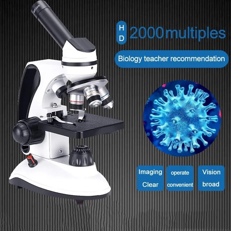
2、 Magnification: Enlarging the image of bacteria for better visibility.
The power of a microscope required to see bacteria depends on the size of the bacteria and the level of detail one wishes to observe. Bacteria are generally very small, ranging from 0.2 to 10 micrometers in size. To visualize such tiny organisms, a microscope with a high magnification power is necessary.
Magnification refers to the ability of a microscope to enlarge the image of an object. The magnification power of a microscope is determined by the combination of lenses it uses. The objective lens, located near the specimen, provides the initial magnification, while the eyepiece lens further magnifies the image for the observer.
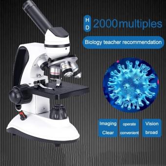
To see bacteria, a compound microscope is typically used. Compound microscopes have multiple lenses that work together to provide high magnification. These microscopes can have various magnification powers, typically ranging from 40x to 1000x or more. The total magnification is calculated by multiplying the magnification of the objective lens by the magnification of the eyepiece lens.
For example, if a microscope has a 10x objective lens and a 10x eyepiece lens, the total magnification would be 100x (10x objective lens multiplied by 10x eyepiece lens). This level of magnification would allow for the visualization of bacteria, but the level of detail may be limited.
To observe bacteria with greater detail, higher magnification powers are required. Microscopes with oil immersion objectives can achieve magnifications of 1000x or more. Oil immersion microscopy involves placing a drop of oil with a refractive index similar to glass between the objective lens and the specimen. This technique minimizes the loss of light and improves resolution, allowing for clearer visualization of bacteria.
It is important to note that while high magnification powers are necessary to see bacteria, magnification alone is not sufficient to observe the fine details of bacterial structures. To visualize the internal structures of bacteria, staining techniques such as Gram staining or fluorescent dyes are often used.
In recent years, advancements in microscopy technology have allowed for even higher magnification powers and improved resolution. Techniques such as electron microscopy, which uses a beam of electrons instead of light, can achieve magnifications of up to several million times. These advanced techniques have revolutionized our understanding of bacterial structures and have contributed to significant advancements in microbiology.
In conclusion, the power of a microscope required to see bacteria depends on the size of the bacteria and the level of detail one wishes to observe. Magnification is crucial for enlarging the image of bacteria for better visibility. Microscopes with magnification powers ranging from 40x to 1000x or more are commonly used to visualize bacteria. Higher magnification powers, such as those achieved with oil immersion objectives, allow for greater detail. Advancements in microscopy technology continue to push the boundaries of magnification and resolution, enabling scientists to explore the intricate world of bacteria with increasing precision.
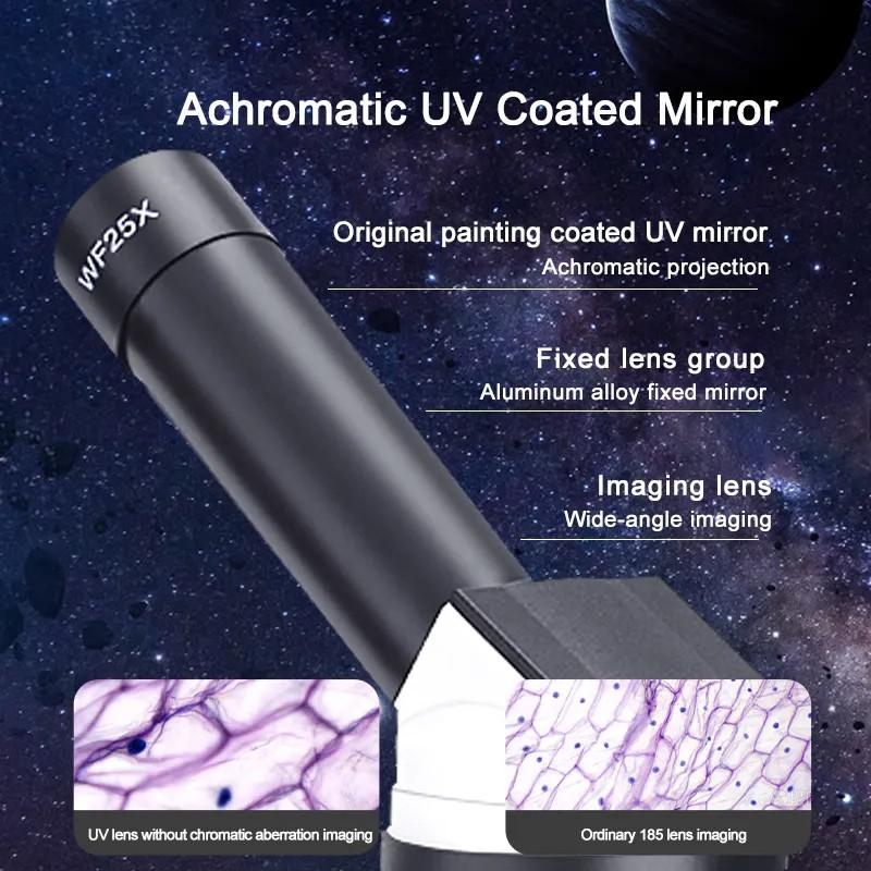
3、 Numerical Aperture: Determines the light-gathering ability of the microscope.
The power of a microscope to see bacteria is determined by its Numerical Aperture (NA). Numerical Aperture is a measure of the light-gathering ability of the microscope, which is crucial for resolving small structures such as bacteria. The higher the NA, the greater the resolving power of the microscope.
To see bacteria, a microscope with a relatively high NA is required. Bacteria are typically in the range of 0.2 to 2 micrometers in size, and to visualize such small structures, a microscope with a high NA is necessary. Microscopes with NA values of 0.65 or higher are commonly used to observe bacteria.
The NA of a microscope is determined by the design of the objective lens and the refractive index of the medium between the lens and the specimen. Higher NA values can be achieved by using immersion oil or other high refractive index media to increase the light-gathering ability of the lens.
It is important to note that while a high NA is necessary for visualizing bacteria, it is not the only factor that determines the quality of the image. Other factors such as the resolution of the microscope, the quality of the lenses, and the lighting conditions also play a significant role.
In recent years, advancements in microscopy techniques have allowed for even higher NA values and improved resolution. Techniques such as super-resolution microscopy and electron microscopy have pushed the boundaries of what can be observed at the bacterial level. These techniques have enabled scientists to study the intricate details of bacterial structures and processes with unprecedented clarity.
In conclusion, a microscope with a high Numerical Aperture is required to see bacteria. The NA determines the light-gathering ability of the microscope, which is crucial for resolving small structures. Advancements in microscopy techniques have further enhanced our ability to visualize bacteria and study their intricate details.
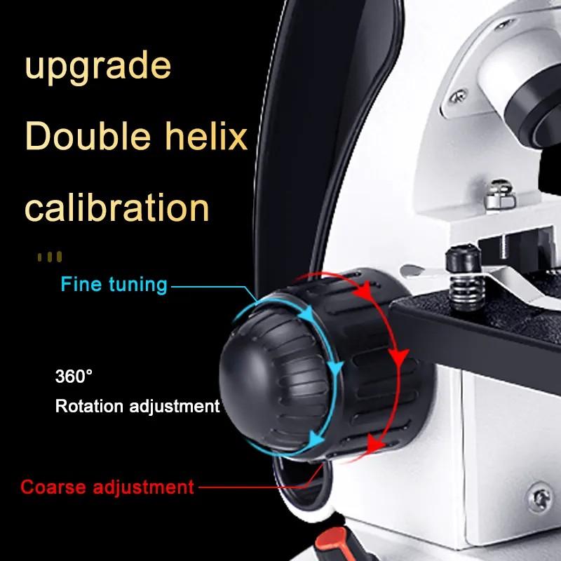
4、 Contrast: Enhancing the differences in color and intensity of bacteria.
To see bacteria under a microscope, a power of at least 1000x is typically required. Bacteria are microscopic organisms that range in size from 0.2 to 10 micrometers, making them too small to be seen with the naked eye. Therefore, a microscope with sufficient magnification is necessary to observe them.
In addition to magnification, contrast is crucial in enhancing the visibility of bacteria. Contrast refers to the ability to distinguish between different elements or structures within a sample. Enhancing the differences in color and intensity of bacteria is one way to improve contrast and make them more visible under the microscope.
There are several techniques that can be used to enhance contrast in bacterial samples. One common method is staining, where specific dyes are applied to the sample to highlight different structures or components of the bacteria. For example, Gram staining is a widely used technique that helps differentiate bacteria into two major groups based on their cell wall composition.
Another technique is phase contrast microscopy, which exploits the differences in refractive index between the bacteria and the surrounding medium. This method allows for the visualization of live, unstained bacteria without the need for additional sample preparation.
In recent years, advancements in microscopy technology have led to the development of new contrast-enhancing techniques. For instance, fluorescence microscopy utilizes fluorescent dyes or proteins to label specific components of the bacteria, allowing for highly specific and sensitive detection.
Overall, a microscope with a power of at least 1000x is necessary to see bacteria. However, enhancing contrast through staining, phase contrast microscopy, or fluorescence microscopy can greatly improve the visibility and detail of bacterial structures, enabling researchers to study them more effectively.










There are no comments for this blog.