What Types Of Microscopes Are There ?
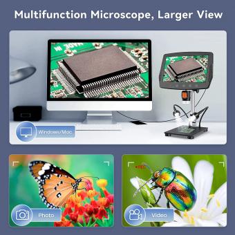
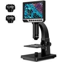
There are several types of microscopes, including optical microscopes, electron microscopes, and scanning probe microscopes. Optical microscopes use visible light to magnify and observe samples. They can be further classified into compound microscopes, stereo microscopes, and digital microscopes. Electron microscopes use a beam of electrons instead of light to achieve higher magnification and resolution. There are two main types of electron microscopes: transmission electron microscopes (TEM) and scanning electron microscopes (SEM). TEMs transmit electrons through a thin sample to create an image, while SEMs scan a focused beam of electrons across the sample's surface. Scanning probe microscopes use a physical probe to scan the surface of a sample and create an image. This category includes atomic force microscopes (AFM) and scanning tunneling microscopes (STM). Each type of microscope has its own advantages and applications in various scientific fields.
1、 Optical Microscopes
Optical microscopes are one of the most commonly used types of microscopes in scientific research and education. They use visible light and a series of lenses to magnify and observe small objects or organisms. Optical microscopes have been instrumental in advancing our understanding of the microscopic world and have been widely used in various fields such as biology, medicine, materials science, and forensics.
There are several types of optical microscopes, each with its own unique features and applications. The most basic type is the compound microscope, which uses two sets of lenses to magnify the specimen. Compound microscopes can achieve magnifications of up to 2000x and are commonly used in biological research and education.
Another type is the stereo microscope, also known as a dissecting microscope. This microscope provides a three-dimensional view of the specimen and is often used for dissection, inspection, and manipulation of larger objects. Stereo microscopes have lower magnification capabilities compared to compound microscopes but offer a wider field of view and greater depth perception.
In recent years, there have been advancements in optical microscopy techniques that have revolutionized the field. One such technique is confocal microscopy, which uses a laser to scan the specimen and create high-resolution, three-dimensional images. Confocal microscopy has enabled researchers to study the internal structures of cells and tissues with exceptional clarity and precision.
Another significant development is the advent of super-resolution microscopy, which surpasses the diffraction limit of light and allows for imaging at the nanoscale. Techniques such as stimulated emission depletion (STED) microscopy and stochastic optical reconstruction microscopy (STORM) have made it possible to visualize structures and processes within cells at unprecedented resolutions.
In conclusion, optical microscopes, including compound and stereo microscopes, have been the workhorses of scientific research for centuries. However, recent advancements in techniques such as confocal microscopy and super-resolution microscopy have expanded the capabilities of optical microscopy, enabling researchers to delve deeper into the microscopic world and uncover new insights.
2、 Electron Microscopes
There are several types of microscopes used in scientific research and various fields of study. One of the most powerful and widely used types is the Electron Microscope (EM). Electron microscopes use a beam of electrons instead of light to magnify the specimen, allowing for much higher resolution and greater detail.
There are two main types of electron microscopes: Transmission Electron Microscopes (TEM) and Scanning Electron Microscopes (SEM). TEMs are used to study the internal structure of specimens by passing a beam of electrons through a thin section of the sample. This produces a high-resolution image that reveals the fine details of the specimen's internal structure. SEMs, on the other hand, are used to study the surface of specimens. They scan the surface with a focused beam of electrons and create a three-dimensional image of the specimen's surface topography.
In recent years, there have been advancements in electron microscopy techniques. One notable development is the introduction of cryo-electron microscopy (cryo-EM). Cryo-EM allows researchers to study biological samples in their native, hydrated state, without the need for staining or fixation. This technique has revolutionized the field of structural biology, enabling the determination of high-resolution structures of complex biomolecules such as proteins and viruses.
Furthermore, there have been advancements in the field of electron tomography, which allows for the reconstruction of three-dimensional structures from a series of two-dimensional images. This technique has been instrumental in studying the architecture of cells and organelles in great detail.
In conclusion, electron microscopes, including TEMs and SEMs, have been instrumental in advancing our understanding of the microscopic world. Recent advancements in electron microscopy techniques, such as cryo-EM and electron tomography, have further expanded the capabilities of electron microscopy and opened up new avenues for scientific exploration.
3、 Scanning Probe Microscopes
Scanning Probe Microscopes (SPMs) are a type of microscope that allows scientists to observe and manipulate materials at the nanoscale level. They are widely used in various fields of science and technology, including physics, chemistry, biology, and materials science. SPMs provide high-resolution imaging and characterization of surfaces, enabling researchers to study the properties and behavior of individual atoms and molecules.
There are several types of Scanning Probe Microscopes, each with its own unique capabilities and applications. The most common types include:
1. Atomic Force Microscope (AFM): AFM uses a small probe with a sharp tip to scan the surface of a sample. It measures the forces between the tip and the sample, allowing for the creation of a three-dimensional image of the surface topography. AFM can also be used to measure various properties such as surface roughness, mechanical properties, and electrical conductivity.
2. Scanning Tunneling Microscope (STM): STM operates by scanning a conducting probe very close to the surface of a sample. It measures the tunneling current that flows between the probe and the sample, providing atomic-scale resolution. STM is particularly useful for studying conductive materials and has been instrumental in the field of nanotechnology.
3. Magnetic Force Microscope (MFM): MFM is a specialized type of AFM that measures the magnetic forces between the tip and the sample. It can map the magnetic field distribution of a sample with high spatial resolution, making it valuable for studying magnetic materials and devices.
4. Electrochemical Scanning Probe Microscope (EC-SPM): EC-SPM combines the capabilities of AFM with electrochemical measurements. It allows researchers to study electrochemical processes at the nanoscale, such as corrosion, electrodeposition, and battery performance.
In recent years, there have been advancements in SPM technology, leading to the development of new techniques and variations of these microscopes. For example, high-speed AFM has been developed, enabling real-time imaging of dynamic processes at the nanoscale. Additionally, new modes of operation, such as Kelvin Probe Force Microscopy (KPFM) and Conductive AFM (C-AFM), have been introduced to study electrical properties and charge transport in materials.
Overall, Scanning Probe Microscopes continue to play a crucial role in nanoscience and nanotechnology, providing valuable insights into the behavior of materials at the atomic and molecular level.
4、 Confocal Microscopes
Confocal microscopes are a type of advanced optical microscope that have revolutionized the field of microscopy. They offer several advantages over traditional microscopes, allowing researchers to obtain high-resolution, three-dimensional images of biological samples.
Confocal microscopes use a laser beam to scan the sample, and a pinhole aperture to eliminate out-of-focus light. This technique, known as confocal imaging, enables the microscope to capture images at different depths within the sample, resulting in a sharp and clear image with improved contrast and resolution. This is particularly useful for studying thick specimens, such as tissues or living cells.
There are several types of confocal microscopes available, each with its own unique features and capabilities. The most common type is the laser scanning confocal microscope (LSCM), which uses a single laser beam to scan the sample point by point. Another type is the spinning disk confocal microscope (SDCM), which uses a spinning disk with multiple pinholes to scan the sample in parallel, allowing for faster image acquisition.
In recent years, there have been advancements in confocal microscopy technology. For example, the development of super-resolution confocal microscopy techniques, such as stimulated emission depletion (STED) microscopy and structured illumination microscopy (SIM), has allowed researchers to achieve even higher resolution imaging. These techniques overcome the diffraction limit of light, enabling the visualization of structures at the nanoscale level.
Furthermore, confocal microscopes are now equipped with advanced imaging software that allows for real-time image processing, 3D reconstruction, and analysis of the acquired data. This has greatly facilitated the study of dynamic processes within cells and tissues.
In conclusion, confocal microscopes are a powerful tool in the field of microscopy, offering high-resolution, three-dimensional imaging capabilities. With ongoing advancements in technology, these microscopes continue to play a crucial role in advancing our understanding of biological systems.


