What's A Compound Microscope ?
A compound microscope is a type of microscope that uses multiple lenses to magnify the image of a specimen. It consists of an objective lens close to the specimen and an eyepiece lens near the observer's eye. The objective lens collects light from the specimen and forms a magnified real image, which is further magnified by the eyepiece lens to produce a virtual image that can be seen by the observer. Compound microscopes are commonly used in scientific research, education, and various fields of microscopy. They offer higher magnification and resolution compared to simple microscopes, allowing for detailed examination of small objects or structures.
1、 Optical magnification
A compound microscope is a type of microscope that uses multiple lenses to magnify an object. It is a versatile and widely used tool in scientific research, education, and various industries. The basic design of a compound microscope consists of two optical systems: the objective lens and the eyepiece.
The objective lens is responsible for gathering light from the specimen and forming a magnified image. It is typically a combination of several lenses with different magnification powers. The eyepiece, or ocular lens, further magnifies the image formed by the objective lens and allows the viewer to observe the specimen.
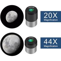
Optical magnification is the primary function of a compound microscope. It refers to the increase in apparent size of an object when viewed through the microscope compared to the naked eye. The magnification power of a compound microscope is determined by the combination of the objective lens and the eyepiece. The total magnification is calculated by multiplying the magnification of the objective lens by the magnification of the eyepiece.
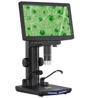
In recent years, advancements in technology have led to the development of digital compound microscopes. These microscopes incorporate digital imaging systems, allowing the captured images to be displayed on a computer screen or other digital devices. This enables easier sharing, analysis, and documentation of the observed specimens.
Furthermore, some modern compound microscopes are equipped with additional features such as phase contrast, fluorescence, or confocal imaging capabilities. These enhancements enable researchers to study specific structures or processes within the specimen with greater detail and precision.
Overall, the compound microscope remains an essential tool in various scientific disciplines, providing researchers and educators with the ability to explore the microscopic world and gain valuable insights into the intricacies of life and matter.

2、 Objective and eyepiece lenses
A compound microscope is a type of microscope that utilizes a system of objective and eyepiece lenses to magnify small objects or specimens. It is one of the most commonly used microscopes in scientific research, education, and various fields of study.
The objective lenses are located at the lower end of the microscope and are responsible for gathering light from the specimen and magnifying it. These lenses are available in different magnification powers, typically ranging from 4x to 100x or higher. The eyepiece lens, on the other hand, is located at the upper end of the microscope and further magnifies the image produced by the objective lenses. The combination of the objective and eyepiece lenses allows for a total magnification of the specimen.
The compound microscope has been a fundamental tool in the field of biology, allowing scientists to observe and study cells, microorganisms, and other small structures in great detail. It has played a crucial role in advancing our understanding of the microscopic world and has contributed to numerous scientific discoveries.
In recent years, advancements in technology have led to the development of more sophisticated compound microscopes. These include features such as digital imaging capabilities, allowing for the capture and analysis of images and videos. Additionally, there have been improvements in the quality of lenses, resulting in higher resolution and clearer images.
Overall, the compound microscope remains an essential tool in various scientific disciplines, enabling researchers and students to explore the intricate details of the microscopic world and uncover new insights into the workings of life.

3、 Illumination system
A compound microscope is a type of microscope that uses multiple lenses to magnify small objects or specimens. It is called a compound microscope because it utilizes two sets of lenses - the objective lens and the eyepiece lens - to provide a higher level of magnification than a simple microscope.
The objective lens is located near the specimen and is responsible for gathering light and forming the primary image of the specimen. It typically has multiple lenses with different magnification powers, allowing the user to switch between different levels of magnification. The eyepiece lens, on the other hand, is located near the user's eye and further magnifies the image formed by the objective lens, making it easier to view.
One crucial component of a compound microscope is the illumination system. This system provides the necessary light to illuminate the specimen, allowing it to be seen clearly. In most modern compound microscopes, the illumination system consists of a light source, such as an LED or halogen lamp, which is directed towards the specimen through a condenser lens. The condenser lens focuses the light onto the specimen, enhancing the contrast and visibility of the image.
The latest advancements in compound microscopes have seen the integration of digital imaging technology. Many microscopes now come equipped with built-in cameras or the ability to attach external cameras, allowing users to capture images and videos of the specimens they are observing. This digital integration has revolutionized the field of microscopy, enabling researchers to easily document and share their findings.
In conclusion, a compound microscope is a powerful tool that utilizes multiple lenses to magnify small objects or specimens. Its illumination system, along with recent advancements in digital imaging, has made it an indispensable instrument in various scientific fields, including biology, medicine, and materials science.

4、 Resolution and image quality
A compound microscope is a type of microscope that uses multiple lenses to magnify small objects or specimens. It consists of an objective lens, which is closest to the specimen, and an eyepiece lens, which is closest to the viewer's eye. The objective lens collects light from the specimen and forms a magnified image, which is then further magnified by the eyepiece lens for the viewer to see.
Resolution and image quality are crucial aspects of a compound microscope. Resolution refers to the ability of the microscope to distinguish between two closely spaced objects as separate entities. It is determined by the numerical aperture of the objective lens and the wavelength of light used. The higher the numerical aperture and the shorter the wavelength, the better the resolution. This allows for clearer and more detailed images of the specimen.
Image quality is influenced by various factors, including the quality of the lenses, the lighting conditions, and the specimen preparation techniques. High-quality lenses with minimal aberrations and distortions contribute to better image quality. Adequate lighting, such as using a bright and uniform light source, ensures optimal illumination of the specimen. Proper specimen preparation techniques, such as staining or mounting, can enhance contrast and improve image quality.
In recent years, advancements in technology have led to the development of digital compound microscopes. These microscopes incorporate digital cameras and software, allowing for real-time imaging, image capture, and analysis. This has revolutionized microscopy by enabling researchers to easily document and share their findings, as well as perform quantitative analysis on the captured images.
Overall, the compound microscope remains a fundamental tool in various scientific fields, including biology, medicine, and materials science. Its ability to provide high-resolution and high-quality images continues to contribute to our understanding of the microscopic world.


