What's The Highest Magnification On A Microscope ?
The highest magnification on a light microscope is typically around 1000x. However, with the use of additional lenses and techniques such as oil immersion, it is possible to achieve magnifications up to 2000x or higher. Electron microscopes, on the other hand, can achieve much higher magnifications, ranging from thousands to millions of times.
1、 Optical Microscopes: Up to 2000x magnification.
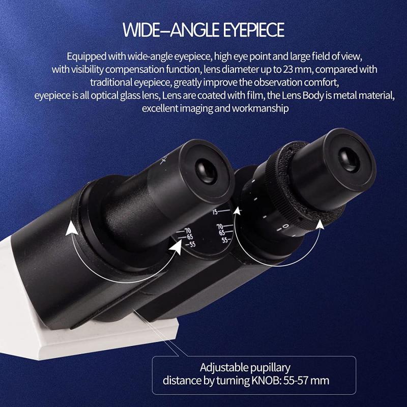
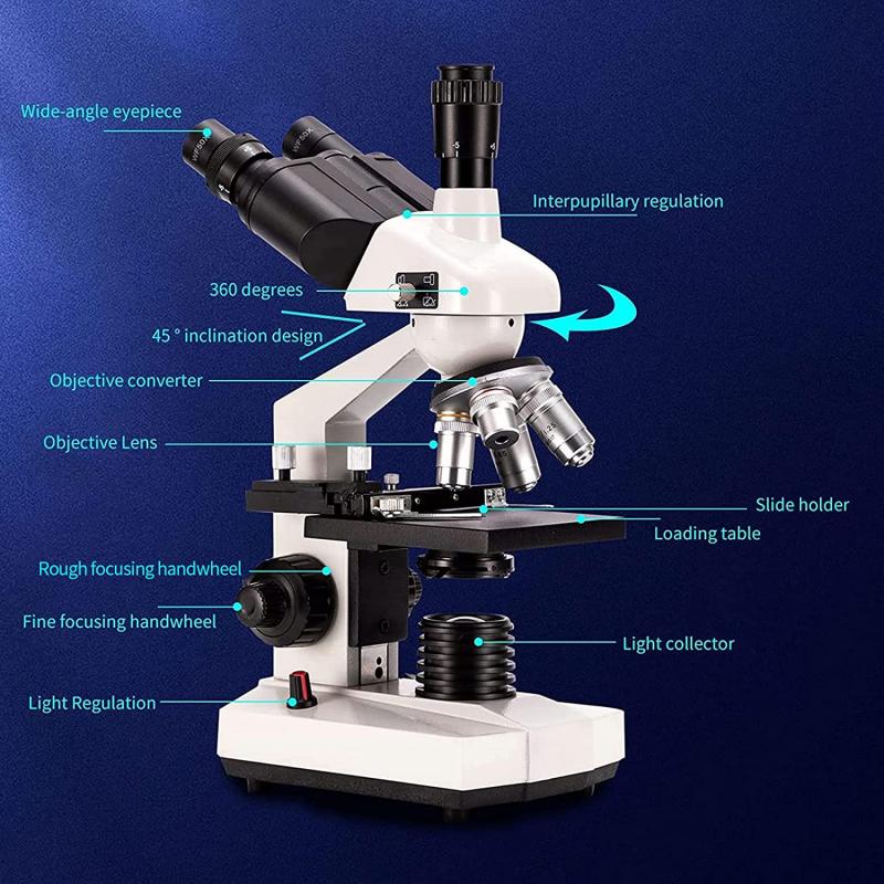
The highest magnification on an optical microscope is typically up to 2000x. Optical microscopes use visible light to magnify and observe specimens. They consist of a series of lenses that work together to increase the size of the image. The magnification power of a microscope is determined by the combination of the objective lens and the eyepiece.
The objective lens is the primary magnifying lens and is available in various magnification powers, typically ranging from 4x to 100x. The eyepiece, or ocular lens, further magnifies the image produced by the objective lens. Most microscopes have eyepieces with a magnification power of 10x. By multiplying the magnification of the objective lens by the magnification of the eyepiece, the total magnification of the microscope is determined.
While a maximum magnification of 2000x is commonly stated for optical microscopes, it is important to note that achieving this level of magnification may require the use of oil immersion techniques. Oil immersion is a method where a drop of oil with a refractive index similar to that of the glass slide is placed between the objective lens and the specimen. This technique allows for higher resolution and magnification.
It is worth mentioning that advancements in technology have led to the development of more specialized microscopes with higher magnification capabilities. For example, electron microscopes can achieve magnifications of up to several million times, allowing for the observation of extremely small structures at the nanoscale. However, these types of microscopes operate on different principles and are not considered optical microscopes.
In summary, the highest magnification on an optical microscope is typically up to 2000x, but achieving this level of magnification may require the use of oil immersion techniques.
2、 Electron Microscopes: Up to 50,000x magnification.
The highest magnification on a microscope depends on the type of microscope being used. Traditional light microscopes, which use visible light to magnify objects, typically have a maximum magnification of around 2000x. However, when it comes to electron microscopes, the magnification capabilities are significantly higher.
Electron microscopes use a beam of electrons instead of light to magnify objects, allowing for much higher resolution and magnification. There are two main types of electron microscopes: transmission electron microscopes (TEM) and scanning electron microscopes (SEM).
Transmission electron microscopes can achieve magnifications of up to 50,000x or even higher. These microscopes use a thin sample that is illuminated by a beam of electrons, and the resulting image is formed by the electrons that pass through the sample. This allows for extremely detailed imaging of the internal structure of cells and other small objects.
Scanning electron microscopes, on the other hand, can achieve magnifications of up to around 100,000x. These microscopes use a focused beam of electrons to scan the surface of a sample, creating a three-dimensional image. This type of microscope is particularly useful for studying the surface features of objects, such as the texture of a material or the shape of a cell.
It is important to note that while electron microscopes offer incredibly high magnification, they also have limitations. For example, electron microscopes require a vacuum environment, which means that living specimens cannot be observed directly. Additionally, the preparation of samples for electron microscopy can be complex and time-consuming.
In recent years, there have been advancements in microscopy techniques that have pushed the boundaries of magnification even further. For example, researchers have developed super-resolution microscopy techniques that can achieve magnifications beyond the diffraction limit of light. These techniques, such as stimulated emission depletion (STED) microscopy and structured illumination microscopy (SIM), have allowed scientists to visualize structures at the nanoscale level.
In conclusion, the highest magnification on a microscope depends on the type of microscope being used. Electron microscopes, specifically transmission electron microscopes and scanning electron microscopes, can achieve magnifications of up to 50,000x and 100,000x, respectively. However, advancements in microscopy techniques continue to push the boundaries of magnification, allowing for even higher resolution imaging at the nanoscale level.
3、 Scanning Probe Microscopes: Up to 100,000x magnification.
The highest magnification on a microscope depends on the type of microscope being used. Traditional light microscopes typically have a maximum magnification of around 1000x. However, when it comes to Scanning Probe Microscopes (SPMs), the magnification capabilities are significantly higher.
Scanning Probe Microscopes, such as Atomic Force Microscopes (AFMs) and Scanning Tunneling Microscopes (STMs), offer exceptional magnification capabilities that surpass those of traditional light microscopes. These microscopes utilize a different imaging technique that allows for higher resolution and magnification.
AFMs, for example, can achieve magnifications of up to 100,000x. They work by scanning a tiny probe over the surface of a sample, measuring the forces between the probe and the sample to create a detailed image. This technique allows for the visualization of atomic and molecular structures with remarkable precision.
It is important to note that the magnification alone does not determine the quality of the image. Factors such as resolution, contrast, and the type of sample being observed also play a crucial role. Additionally, advancements in technology continue to push the boundaries of microscope magnification capabilities.
In recent years, there have been significant developments in microscopy techniques, such as super-resolution microscopy, which have allowed for even higher magnification and resolution. These techniques utilize various methods, such as structured illumination and single-molecule localization, to surpass the diffraction limit of light and achieve resolutions beyond what was previously thought possible.
In conclusion, the highest magnification on a microscope depends on the type of microscope being used. Scanning Probe Microscopes, such as AFMs, can achieve magnifications of up to 100,000x, allowing for the visualization of atomic and molecular structures. However, it is important to consider other factors such as resolution and sample type when evaluating the quality of the image. Ongoing advancements in microscopy techniques continue to push the boundaries of magnification capabilities, enabling researchers to explore the microscopic world with unprecedented detail.
4、 Super-resolution Microscopy: Beyond the diffraction limit.
The highest magnification on a microscope is not solely determined by the microscope itself, but also by the technique used for imaging. Traditional light microscopy is limited by the diffraction of light, which restricts the resolution to about half the wavelength of light (around 200-300 nanometers). However, advancements in microscopy techniques have allowed scientists to overcome this diffraction limit and achieve super-resolution imaging.
Super-resolution microscopy techniques, such as stimulated emission depletion (STED) microscopy, structured illumination microscopy (SIM), and single-molecule localization microscopy (SMLM), have revolutionized the field of microscopy by enabling imaging at resolutions beyond the diffraction limit.
STED microscopy uses a combination of laser beams to excite and deplete fluorescent molecules, resulting in a highly confined focal spot that can be smaller than the diffraction limit. This technique has achieved resolutions down to a few tens of nanometers.
SIM utilizes patterned illumination to extract high-frequency information from the sample, allowing for resolution improvements of up to two times the diffraction limit. This technique has been used to visualize cellular structures with resolutions of around 100 nanometers.
SMLM, including techniques like stochastic optical reconstruction microscopy (STORM) and photoactivated localization microscopy (PALM), relies on the precise localization of individual fluorescent molecules. By sequentially activating and localizing sparse subsets of molecules, super-resolution images with resolutions of a few nanometers have been achieved.
It is important to note that the highest achievable magnification on a microscope depends on various factors, including the numerical aperture of the objective lens, the quality of the optics, and the sample itself. However, with super-resolution microscopy techniques, researchers can now visualize and study biological structures and processes at unprecedented resolutions, providing valuable insights into the nanoscale world.
